Reports: UR552145-UR5: Synthesis of Ruthenium Dioxide Nanoparticles and Clusters with Tailored Size-and Surface-Dependent Properties for Enhanced Catalytic Applications

Vladimir Kitaev, Wilfrid Laurier University
In the final year of the project with the remaining finding, we have concentrated on the preparation of iron oxide and oxyhydroxide nanoparticles, specifically: 1) size control in rhombohedral hematite nanoparticles; 2) 2-D morphologies of iron oxides and oxyhydroxides; and 3) porous iron oxide nanoparticles. The main interest in iron nanoparticles is that they are purely inorganic-based nanoparticle systems, the synthesis of which can be scaled to multi-grams (or even kgs) of nanoparticles, as well iron nanoparticles alone gave very promising results in degradation and removal of organophosphorous compounds by chemical and photochemical treatment.

1.  Size control in rhombohedral hematite nanoparticles. 

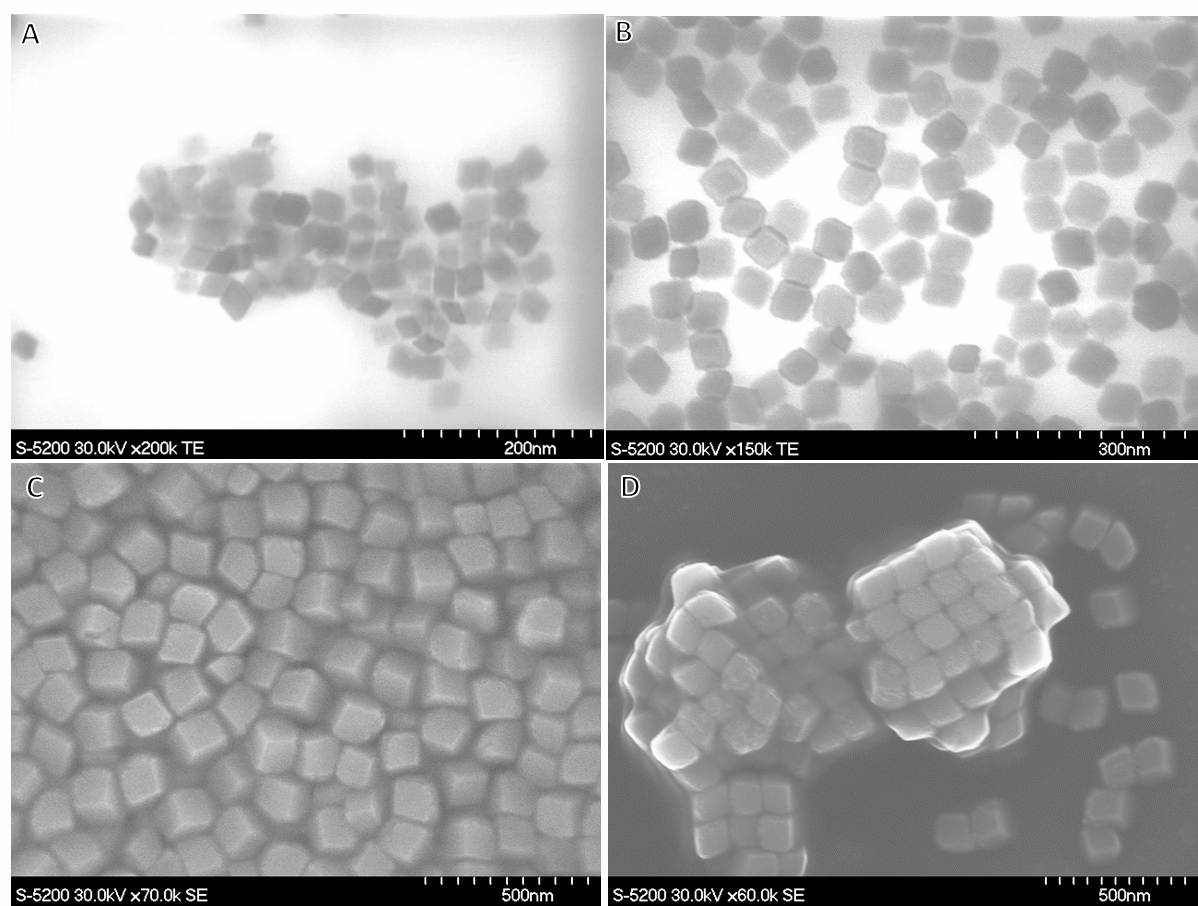
Figure 1. Electron microscopy images of rhombohedral hematite nanoparticles with average sizes of A) 35 nm, B) 50 nm, C) 100 nm, and D) 120 nm with the self-assembled arrays shown.

Rhombohedral hematite nanoparticles with particle sizes from ca. 30 nm to 200 nm were prepared (Figure 1). In several consecutive iterations of the synthesis, nanoparticles size dispersity was improved from ca. 15-20% to 5-10% in standard deviation of the size. Polydispersity index (PDI) measured with the Zetasizer is in the range of 0.025 to 0.035. Synthesised hematite particles with low size dispersity show self-assembly (Figure 1D), which can be advantageous for preparation of porous oxide materials and photonic materials based on colloidal photonic crystals.

In addition to electron microscopy, shown in Figure 1, hematite nanoparticles were characterized by XRD (with the pure hematite phase confirmed), Zetasizer to estimate the size, size dispersity and charge in solution, and UV-vis spectroscopy. The next stage in this project is to work on accessing hematite seeds of smaller sizes than current 30 nm and to prepare larger nanoparticles by their seeded regrowth.

2.  2-D morphologies of iron oxides and oxyhydroxides.

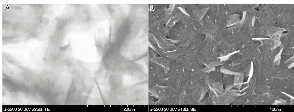
Figure 2. Electron microscopy images of the sheet morphologies of iron oxyhydroxides A) transmission electron microscopy image; B) scanning electron microscopy image.

Sheet-like 2-D morphologies of iron oxyhydroxides were prepared by controlled hydrolysis of iron precursors in combination with degradable organic complexing agents, such as formic and oxalic acid and paraformaldehyde. The thickness of the obtained oxide layers are under 2 nm, which gives very large surface area advantageous for many applications, especially degradation and removal of the pollutants. The average sheet size of several samples (such as shown in Figure 2) is remarkably constant. The polydispersity index (PDI) of the colloidal dispersions of these sheets measured with the Zetasizer is 0.062, which is a very low value for such anisotropic particles. We are currently further exploring this system, especially their conversion to hematite with the preservation of the morphology.

3.  Iron oxide nanoparticles with high porosity.

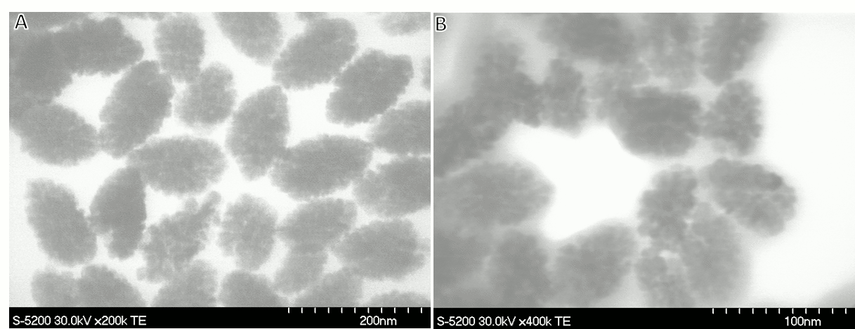
Figure 3. Transmission electron microscopy images of the porous iron oxide nanoparticles A) ellipsoids of ca. 60 nm by 120 nm; B) particles of ca. 30 nm by 60 nm in size.

Since high porosity and large surface area are advantageous for many applications of oxide nanoparticles, we have explored the preparation of porous nanoparticles of iron oxide using controlled hydrolysis in combination with removable organic ligands. Several successful syntheses have achieved smaller particles (ca. 5-10 nm) as a constituent of larger particles of ca. 50-100 nm in size (Figure 3). 50-100 nm is a target size for convenient colloidal deposition of functional monolayers tested previously in this project. The characteristic egg-like shape of these nanoparticles (Figure 3) is inherited from spindle morphologies of akaganeite. The next step in this direction is assuring complete conversion to hematite with the preservation of the porosity.

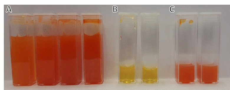
Figure 4. Optical photographs of iron oxide and oxyhydroxide nanoparticle dispersions A) hematite samples representative of images shown in Figure 1; B) oxyhydroxide samples representative of images shown in Figure 2; B) samples of porous iron oxide shown in Figure 3. 

Optical photographs of the prepared nanoparticle samples are shown in Figure 4. Figure 4A presents several hematite samples of different size. The 2-D morphologies of the iron oxyhydroxide are yellow dispersions in Figure 4B. Porous iron oxide nanoparticles are displayed in Figure 4C; these samples are predominantly hematite, as evidenced from the colour of nanoparticle dispersions.

In terms of applications, we will be seeking collaborations in development of iron and manganese oxide as anode materials for water splitting and photoelectrochemical degradation of pollutants in wastewater. Currently, in collaboration with Dr. Scott Smith (Wilfrid Laurier University, joined MSc student Farah Ateeq), we have obtained promising preliminary results on degradation and removal of organophosphorous compounds. We have tested series of manganese and iron oxide nanoparticles with hydrogen peroxide as an oxidant. We found several systems where reasonably small amount of hydrogen peroxide (0.05 M) can be used successfully and that have broad action against all classes of organophosphorous pollutants. This work is now continued with the transfer of the successful removal systems for the treatment of real wastewater samples. 

Overall, the project in its duration achieved preparation of several classes of photoelectrochemically active oxides, starting with ruthenia and iridium oxide and subsequently manganese and iron oxides. Colloidal preparation for different morphologies of oxide nanoparticles has been successfully developed with size and shape selection, including thin 2-D layers and porous particles advantageous for applications. Nanocomposites of metal oxide with metals (e.g. Au&MnO2) have been prepared. Photoelectrochemical properties were extensively tested and applications in degradation of pollutants were successfully initiated.