Reports: DNI753116-DNI7: Atomic-Scale Visualization of Paraffin Melting and Crystallization with Ultrafast Transmission Electron Microscopy
 printer friendly
Having the ACS PRF DNI grant has been instrumental for enabling work that forms the foundation of several projects – including ultrafast imaging of structural dynamics in soft matter – that are now federally funded and supported through industrial collaborations or philanthropic foundations. As predicted in the first-year report, funding from the ACS PRF has very-successfully served as seed money for generating preliminary results on achieving combined high spatial and temporal resolutions [i.e., nanometer and femtosecond (fs)] with the University of Minnesota ultrafast electron microscopy (UEM) lab. The results summarized below currently (and will continue to) form a critical component of grant submissions. Additionally, the funding provided by the ACS PRF has enabled critical support for graduate students early in their scholarly pursuits, free from the worry of limited instrument time, challenges associated with the purchase of supplies, the inability to travel to conferences, etc. Indeed, the positive impact of such funding early in the career of a PI starting a challenging research program cannot be overemphasized; the work that has been enabled by the ACS PRF DNI is certain to have a lasting impact on the group for years to come. What follows are brief summaries of results of the work that has been enabled by funding from the ACS PRF DNI grant. In addition to these synopses, all of which describe published work, there are several other manuscripts that are currently in some stage of the publication process (e.g., submitted, in press, etc.).
Characterization of Fast Photoelectron Packets in Weak and Strong Laser Fields in Ultrafast Electron Microscopy
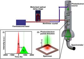
The development of UEM and variants thereof (e.g., photon-induced near-field electron microscopy, PINEM) has made it possible to image nanoscale dynamics on the picosecond timescale (see UEM schematic below). Accessing the fs regime with UEM currently relies on the generation of photoelectrons with an ultrafast laser pulse and operation in a stroboscopic pump-probe fashion. With this approach, temporal resolution is limited mainly by the durations of the pump laser pulse and probe photoelectron packet. The ability to accurately determine the duration of the photoelectron packets, and thus the instrument response function, is critically important for interpretation of dynamics occurring near the temporal resolution limit, in addition to quantifying the effects of the imaging mode. In this work, a technique for in situ characterization of ultrashort photoelectron packets is described; the technique makes use of coupling the photoelectrons with photons in a photoinduced evanescent near-field of the specimen (figure inset). It is shown that within the weakly-interacting (i.e., low pump-laser fluence) regime, the zero-loss peak temporal cross-section is precisely the convolution of electron packet and photon pulse profiles. Beyond this, the effects of non-linear processes are outlined, and it is shown that temporal cross-sections of high-order peaks explicitly reveal the electron packet profile, while use of the zero-loss peak becomes increasingly unreliable.
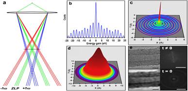
Effects of Quantized Chromatic Aberrations in Ultrafast Electron Microscopy
While the single-photoelectron approach in UEM circumvents deleterious space-charge effects, the narrow energy spread can be compromised when the photon pulses and the electron packets are spatiotemporally overlapped at the specimen; light absorption and emission can occur in the specimen near-field, thus generating large populations of photoelectrons (relative to the elastically-scattered electrons) having discrete energies differing by integer multiples of photon quanta [panel (b)]. This results in a radially symmetric projection of the discrete electron energy distribution in the image plane due to the velocity dependence of the Lorentz force [panel (a)] and, consequently, produces annular chromatic aberrations in the real-space intensity distributions [panels (c) and (d)]. In this work, a description of how the induced electron-energy envelope resulting from absorption and emission of photons in the optically-pumped specimen near-field becomes the dominant effect limiting spatial resolution of UEM images during initial excitation. Moreover, select spatial frequencies become exaggerated due to the quantized nature of the near-field interaction. Application of the effect to representative lattice-fringe images of model nanostructures indeed limits resolving power [panel (e)] and suggests that this transient, quantized chromatic aberration will need to be addressed in order to achieve angstrom-scale, real-space ultrafast imaging with UEM in the initial moments of specimen excitation.
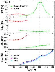
Effects of Thermionic-Gun Parameters on Operating Modes in Ultrafast Electron Microscopy
Ultrafast electron microscopes with thermionic guns and lanthanum hexaboride (LaB6) emission sources can be operated in both the nanosecond, single-shot and fs, single-electron modes. This has been demonstrated with conventional, self-biasing electron guns comprised of a Wehnelt electrode and absent any modifications beyond integration of optical ports into the column. In this work, by conducting simulations using the General Particle Tracer code, the electron-gun parameter space within which various modes (e.g., single-electron per packet) may be optimized is defined. The properties of interest included electron collection efficiency [panel (a)], temporal and energy spreads [panels (b) and (c)], and effects of laser-pulse duration incident on the LaB6 source [panel (d)]. It was found that, depending upon LaB6 position and Wehnelt aperture size, collection efficiencies can reach 100% for all modes, despite there being no bias applied to the electrode. Further, it was found that laser-pulse duration has only a modest effect on the statistical temporal spread of the photoelectron packets. The results suggest there are optimal electron-gun settings for maximum beam current with commensurate effects on temporal and energy spread. Achieving these effects can be accomplished by varying Wehnelt aperture size and LaB6 emission-source position.