Reports: DNI655184-DNI6: Visualizing Molecular Organization and Energy Transport Dynamics at Organic Surfaces and Heterojunctions with Interface Specific Femtosecond Spectroscopy
Sean T. Roberts, PhD, University of Texas at Austin
Organic semiconductors (OSCs) are a
unique class of petroleum-derived materials that blend many of the processing
advantages of plastics with the electrical properties of semiconductors. In
contrast to common semiconductor materials such as silicon and GaAs, OSCs can
be readily processed from solution into highly-absorbing thin, conductive
films, making OSCs attractive materials for use in a wide range of
optoelectronic applications, including photovoltaic cells, light emitting
diodes, and photodetectors. In each of these applications, the transfer of
charge to and from OSCs is fundamental to device operation. The primary
goals of our work carried out through this grant are to (1) develop a
series of interface-specific, nonlinear spectroscopies that can be used to
investigate OSC material interfaces, and (2) establish how the molecular
organization of these regions controls their ability to donate and accept
charge. Specifically, we have been using electronic sum frequency generation, a
nonlinear process that selectively occurs at regions of a sample that
experience a breakage of inversion symmetry, to probe the electronic density of
states of buried OSC interfaces. Following the start of our funding this past
January, we have achieved a series of key milestones for this research project
which are described in detail below:
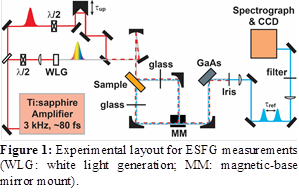
Construction
of an Electronic Sum Frequency Generation (ESFG) Spectrometer:We
have successfully completed the construction of a spectrometer that can be used
to obtain broadband ESFG spectra of thin film samples. Figure 1 illustrates the
design of this instrument. In a broadband ESFG measurement, a small portion of
the output of a Ti:sapphire amplifier is used to generate a supercontinuum
excitation field that spectrally extends from ~400 – 750 nm. This field is
mixed in a target sample with a portion of the 800 nm output from the
Ti:sapphire amplifier, generating an ESFG field in the ultraviolet range that
is detected using a spectrometer and a silicon CCD. Representative ESFG spectra
obtained from organic thin film samples appears in Figures 2 and 3 below. Our
spectrometer is designed such that we can rapidly switch between collecting of
ESFG spectra reflected from a sample surface or transmitted through a sample.
As we describe below, for thin films both transmitted and reflected ESFG
signals can substantially aid in isolating the portion of the signal that
originates from a specific buried interface of interest within a sample. We are
currently working to implement a heterodyne detection scheme that can boost the
strength of our collected ESFG signal by interfering it with a known reference
field generated by refocusing the white light and 800 nm excitation fields onto
a piece of GaAs after the sample. We expect that this will boost our ESFG
signal strength by 10-100×, which will significantly aid in implementing
time-resolved measurements that detect transient changes in the ESFG signal of
a sample following photoexcitation by a femtosecond pump pulse.
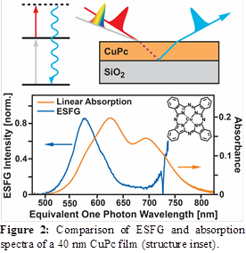
Characterization
of the Interfacial Density of States of Copper Phthalocyanine
Thin Films:As an initial system for study, we
have focused on using ESFG to characterize the interfacial density of states of
copper Phthalocyanine (CuPc) thin films deposited on SiO2. CuPc is
an exceptionally well-studied OSC, making it a fantastic model system for
benchmarking our ESFG spectrometer. Figure 2 plots a comparison between the
bulk absorption spectrum of a 40 nm thick CuPc film and its ESFG response. The
ESFG spectrum is clearly shifted to higher energy. While this result in part
reflects interference between ESFG signals emitted from the exposed CuPc:air
and buried CuPc:SiO2 interface, a fit to our spectral data that
accounts for this interference (see below) suggests that the CuPc HOMO-to-LUMO
transition at the buried interface is spectrally narrowed and shifted to higher
energy at the buried CuPc:SiO2 interface. Such narrowing and
shifting of this transition may reflect both a decrease in excitonic coupling
between CuPc molecules at the buried interface as well as the difference in
dielectric constant between CuPc and SiO2. A manuscript describing
these results is currently in preparation for submission to the Journal of
Physical Chemistry Letters.
In addition to our work on CuPc
films, we have also started preparing thin films of squaraine dyes. These
materials are strong absorbers in the visible and near-infrared spectral range,
but it is unclear how the packing arrangements that these materials adopt at
interfaces in electrical devices affects their ability to accept and transfer
charge. Squaraine dyes for this work are currently being provided to us through
a collaboration with Prof. Mark Thompson’s research group at the University of
Southern California.
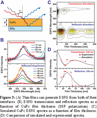
Implementation
of a Thin Film Interference Model for Extracting Signals from Buried
Interfaces: A complication that must be
accounted for when investigating thin films using ESFG is the fact that the
film has two potentially active ESFG interfaces. Given that the thicknesses of
the films that we investigate are on the order of ~50 - 100 nm, it is not
possible to spatially isolate these signals. However, at a detector, these two
signals can interfere with one another. As the thickness of the film is
changed, the phase difference between the ESFG signals emitted from its top and
bottom interfaces can change from destructive to constructive. This causes the
amplitude of the ESFG signal to oscillate with film thickness, providing data
that can be fit to isolate the ESFG response of the buried interface. Over the
course of the past year, we have developed a software package that uses a thin
film transfer matrix modeling approach to calculate the amplitude of the two
driving fields that stimulate ESFG from the sample at each spatial position
within the film. This code uses this information to calculate the emitted ESFG
signal. By fitting how the strength of the emitted signal varies as a function
of film thickness in both reflected and transmitted ESFG spectra, we can
isolate the ESFG response of the buried interface of interest.
 printer friendly