Reports: ND554149-ND5: Impact of Microenvironment on the Catalytic Activity of Supported Gold Nanoclusters
Jennifer Shumaker-Parry, PhD, University of Utah
Ilya Zharov, PhD, University of Utah
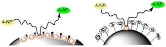
The goal of the proposed research is to
generate controllable microenvironments for silica-supported gold nanoclusters
(AuNCs) and to investigate the impact of the
microenvironment on catalysis (Figure 1).Initially, we worked on exploring the effect of nanoenvironment on the catalytic properties of gold
nanoparticles (AuNPs) immobilized on larger silica
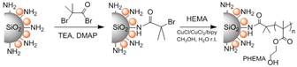
nanoparticles (SiNPs). We prepared aminated SiNPs and deposited ~10 nm AuNPs
on the surface. Next, we alkylated the surface primary amines using 2-bromoisobutyryl bromide, and used
this moiety to initiate atom transfer radical polymerization of 2-hydroxyethyl
methacrylate (Figure 2). This provided polymer brush-grafted nanoparticles
whose polymer brush length was controlled in the range of 5-30 nm by the polymerization
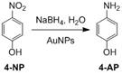
time. We examined the catalytic activity of these nanoparticles using a model
reaction, reduction of 4-nitrophenol (4-NP) to 4 aminophenol (4-AP) with NaBH4
(see below). While we noted clear indications that the polymer brush
length and the presence of hydroxyl groups in the polymer side chain affect the
catalytic activity of AuNPs, we also observed loss of
catalytic activity due to AuNP aggregation because of
rapid dissolution of SiNP supports under the highly
basic reaction conditions (Figure 3). As the result, we shifted our focus to ND
as a more stable catalyst support material.
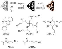
We used thiol-ene chemistry for
both attachment of a polymer film to ND and subsequent immobilization of noble
metal nanoparticles on the ND surface. We used a
photo-initiator 2,2-dimethoxy-2-phenylacetophenone
(DMPA) that was exposed to UV light at 254 nm, generating free radicals that
cleave the S-H bonds in entaerythritol
tetra(3-mercaptopropionate), PETMP, which then combine with vinyl monomers
present in solution and with the double bonds present on the ND surface (Figure
4). Upon treatment with sodium borohydride (NaBH4), Au, Pt, and Pd nanoparticles tethered to the surface of the
polymer-coated NDs were formed, depending on the metal salt present. Figure 5 presents
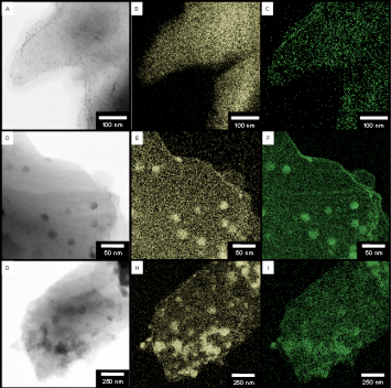
TEM images of Au, Pt and Pd nanoparticles on
polymer-coated ND supports. A closer inspection of these NDs (Figure 5A) shows AuNPs with a diameter of ~3 nm on the polymer/diamond
surface. The AuNPs follow the contour of the diamond
support closely, suggesting that the polymer adhesion layer is thin. In addition to gold, in situ particle growth worked for the formation of Pt and Pd NPs on the ND particles, as can be seen in Figures 5D,G.
High resolution energy dispersive spectroscopy (EDS)
obtained with scanning TEM (S/TEM) provided nanoscale chemical mapping of the
polymer and Au, Pt, or Pd NPs attached to the
polymer/ND surface. As evident from Figure 5, the polymer coating appears to
cover the entire ND surface. The BF-S/TEM images in Figure 5A,D,G
are clearly correlated to the maps of the characteristic sulfur-K X-rays at 2.3
keV shown in Figure 5B,E,H. One of the monomers in
the polymer coating is PETMP which is a tetra-thiol molecule, the only
sulfur-containing molecule. Therefore, the sulfur EDS map indicates that the
polymer coating is confined to the surface of the ND shown in the BF-S/TEM
images. Figure 5C,F,I correspond to the Au-L, Pt-M,
and Pd-L x-ray lines at 9.7, 2.0, and 2.8 keV, respectively. These EDS maps demonstrate that metallic
NPs are adhered to the surface of the thiol-ene
polymer which coats the entire surface of the ND and follows the topography of
the diamond surface with high fidelity. These maps complement the XPS data and
show that the sulfur bonding environment corresponds to the local attachment of
the metallic NPs.
We carried out preliminary studies of the catalytic
efficiencies of the Au, Pt, and Pd NPs supported on
the polymer/ND particles without addition of polymer brushes. We probed the reduction
of 4-NP to 4-AP with NaBH4 (Figure 6), which is easily monitored
using UV-Vis absorbance spectroscopy when this reaction is catalyzed by metal
nanoparticles. Instead of turnover frequencies (TOFs), we quantify the
catalytic activity in units of catalytic cycles per active site per second. When
used as the catalyst for the reduction of 4-NP the STYs for Au, Pt and Pd NPs immobilized on polymer/NDs were observed to be 0.003
± 0.001, 0.021 ± 0.003, and 0.018 ± 0.04 s-1, respectively. Reported
values for Au and Pd nanoparticles in solution follow
a similar trend, with AuNPs being less catalytically
active than Pd when used in the reduction of 4-NP.
However, the activity of the PtNP/polymer/NDs is
higher than expected and it is possible that the attachment of the PtNPs to the polymer may influence the adsorption energy of
4-NP leading to a more favorable reaction environment than that of unsupported PtNPs.
Figure
1.Schematic representation of the proposed idea.
Figure
2. Preparation of poly(2-hydroxyethyl
methacrylate) brushes on silica nanoparticle surface.
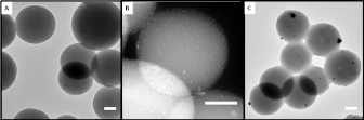
Figure 3. (A) TEM image of AuNP decorated silica spheres
with (B) a higher magnification HAADF-S/TEM image. (C) Silica dissolution and AuNP aggregation after brief exposure to basic solution. All
scale bars are 50 nm.
Figure 4.Photoinitiator
and monomers used in polymer coating of NDs. The latter were then decorated
with metal nanoparticles via in situ
reduction of metal salts.
Figure 5.Bright field (BF-S/TEM) images of polymer/NDs decorated
with (a) Au, (d) Pt, (g) Pd. EDS images extracted from the sulfur K-line of the
same polymer/NDs as seen in the BF-S/TEM images decorated with (b) Au, (e) Pt,
and (h) Pd NPs, respectively. EDS images extracted
from the (c) Au M-line, (f) Pt M-line, (i) Pd L-line
of NP decorated polymer/NDs are of the same regions as seen in the BF-S/TEM and
sulfur K-line EDS images.
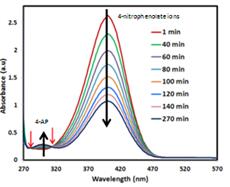
Figure 6. Reaction scheme of
conversion of 4-nitrophenol (4-NP) to 4-aminophenol (4-AP) catalyzed by AuNPs (top). UV-visible absorption
spectra showing conversion of 4-nitrophenol to 4-aminophenol in the presence of
ND-supported AuNPs. Red arrows indicate isosbestic points (bottom).
 printer friendly