Reports: DNI654407-DNI6: Direct Visualization of Interfacial Energy-Transfer and Charge-Transfer Dynamics of Thin Ionic Liquid Films by Ultrafast Electron Imaging
Ding-Shyue Yang, PhD, University of Houston
Introduction
In the last
two decades, ionic liquids (ILs) have attracted great interest in various
research communities and industries due to their remarkable physicochemical properties,
including low melting points, high thermal and electrochemical stability, high
ionic conductivity, hightuneability
as designer solvents, negligible vapor pressure, etc. Much effort has been made
to understand their behaviors in homogeneous liquid and solution phases.
Recently, more and more attention has been drawn to the structures and dynamics
of ILs in a heterogeneous environment because of IL applications in, e.g.,
(photo)catalysis, electrochemistry, corrosion
inhibition, and thin-film lubrication. To study solid-IL interfaces and obtain
the corresponding signals instead of the bulk ones, it is preferred to use experimental
methods with a surface-specific probe that has proper spatiotemporal
resolutions.
Many of the
interfacial studies were conducted using sum frequency generation (SFG), x-ray
reflectivity, x-ray photoelectron spectroscopy, atomic force microscopy (AFM),
and scanning tunneling microscopy (STM). Essentially all of them were time-averaged
measurements, providing a static picture of the solid-IL interfaces in
equilibrium states despite that the relevant time scales for molecular motions
are many orders of magnitude faster. In recent years, ultrathin films of ILs have
be prepared via physical vapor deposition and studied under ultrahigh vacuum
conditions, thanks to the nonvolatile property of ILs at room temperature. However,
a considerable debate about the ion organization and layered structures still exists
in the experimental literature. This is likely due to the different inherent
challenges encountered in each technique regarding the probing of interfacial
structures: force-dependent observation in AFM, ion adsorption on the tip in
STM, beam-induced damage by long exposure of x-ray, and ensemble averaging
using optical techniques.
Our method of
choice is time-resolved electron diffraction to study the structures and
ultrafast dynamics at solid-IL interfaces. Compared to x-ray photons, electrons
have orders-of-magnitude higher scattering cross sections with matter and can
be easily generated (thermally or via photoemission) and manipulated using
electron optics. These advantages make electrons a natural choice of contact-free probe in interfacial
studies, with surface specificity and direct
probing of the structures of molecular assemblies. Our research plan has two
main goals: (1) to examine the
structural orders of IL thin films supported on various types of substrate
surfaces at different temperatures, and (2)
to obtain information of photoinduced energy-transfer and charge-transfer
dynamics across solid-IL interfaces. We focused on the first goal during the
first year of the project.
Results and Discussion
The
representative IL of 1-ethyl-3-methylimidazolium bis(trifluoromethanesulfonyl)amide, [emim][NTf2],
was chosen based on the consideration of thermal stability for vapor deposition
and the comparable sizes of cations, [emim]+,
and anions, [NTf2]–, for potentially better packing
orders. A Knudsen-type effusion cell holding the IL was heated to around 430 K
for the deposition of thin films, whose rate has been calibrated using a quartz
crystal microbalance directly under the cell output. By changing the deposition
time, IL films of ~1 up to 20 nanometer (nm) thickness were deposited on
different substrate surfaces, including highly-oriented pyrolytic graphite
(HOPG), hydrogen-terminated Si(111), mica, Cu(111),
and Ni(111).
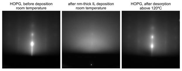
Figure 1
shows the diffraction images of a solid-IL interface with and without IL
coverage, using HOPG as an example. An adsorbate-free substrate surface can be
recovered after desorption of the IL thin film at an elevated temperature (see Fig.
1, left and right images).
We found that
all of the nm-thick films deposited on substrates held at room temperature give
a diffuse scattering pattern without clear diffraction spots or rings (the
middle image of Fig. 1). Such an observation signifies the poor vertical and
horizontal, or local, ordering of cations and anions at the solid-IL interface
when no bias voltage is applied. The best description for the structure of these
IL thin films is a liquid-like
configuration with no apparent nm-sized crystalline order.
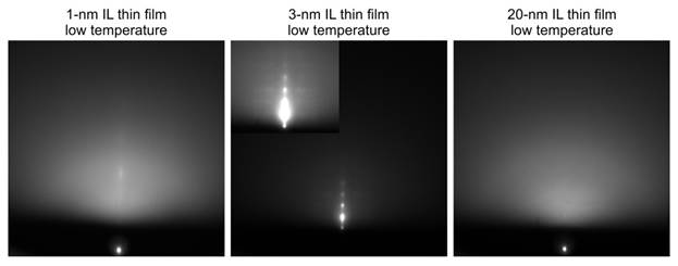
Following the
deposition, the structures at the solid-IL interface were monitored at
different temperatures. It is noted that, among all of the substrates studied
so far, only ~3-nm-thick IL films on HOPG repeatedly show clear Bragg
diffraction spots as the temperature decreases to ~30 degrees below the IL melting
point (Figure 2, the middle image and the intensity-boosted inset). Interestingly,
films of much smaller and much larger thickness on HOPG do not produce such temperature-dependent
diffraction changes (Fig. 2, left and right images). Based on simulations using
the kinematic scattering theory, we confirmed that the Bragg diffractions come
from crystals of the IL well-ordered in the surface normal direction but
azimuthally rotated in the horizontal two dimensions. Cations and anions in the
thin film are packed in the checkboard-type arrangement following the structure
of the bulk crystal. Moreover, the vertical order originates from the lattice
matching between the IL’s c-axis cell
constant and graphite’s interplanar distance. Thus, due
to its surface steps and terrace, HOPG serves as a template to promote the
vertical order in the 3-nm thin film. It is also consistent that this template
effect is not pronounced in a 1-nm-thick film, and
ions in a much thicker film farther away from the stepped surface yield a
liquid-like arrangement without clear crystalline order.
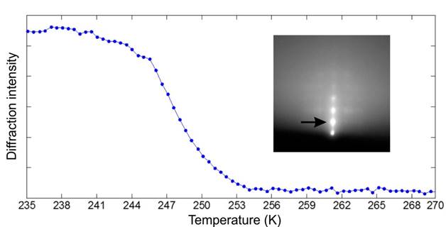
By following
the intensities of the Bragg diffractions, we found that the loss of the
crystalline order in the IL thin film starts below, but completes at, the bulk
melting temperature of 252 K (see Figure 3). Such an observation signifies that,
with the assistance of a template, the IL thin film behaves similarly to the
bulk. This is consistent with the picture that the Coulomb interactions between
ions are the driving force in the behavior of the IL.
Impact and Future Plans
These new diffraction
results show intriguing solid-IL interfacial interactions on the nanometer
scale and warrant more studies of structures and dynamics. The proposed
research will broaden the applications of ultrafast electron diffraction in
chemistry and condensed matter, which is a major focus for us. Three graduate students,
KarjiniRajagopal, Chengyi Wu, and NapatPunpongjareorn, has participated in this project and
acquired direct experiences working with ILs and advanced experimental
techniques. We are preparing a manuscript for this work.
 printer friendly