Reports: DNI653820-DNI6: Catalytic Activity of Functionalized Graphene: Influence of Charge Carrier Dynamics
 printer friendly
Catalysts and photo-catalysts are widely applied in systems related to generating, and converting energy, such as oil refinery, new fuel productions, and are also discussed, e.g. for petroleum refinery wastewaters treatment. Charge carrier transfer reactions are fundamental for catalytic and photo-catalytic processes. Catalysis in general involves charge transport or charge redistribution, followed by a change in the potential energy landscape, and subsequent bond breaking and/or bond formation processes on the femto- to picosecond time-scale. Understanding charge carrier dynamics in materials for heterogeneous catalysis is a key requisite for developing and improving catalysts and photo-catalysts.
The objectives of our project are the investigation of mechanisms leading to an enhancement of photo-catalytic activity. Charge carrier mobility at the surface and charge carrier trap state density are important factors for heterogeneous catalysis at interfaces. Metal oxides constitute a large class of catalysts. The surfaces of metal oxide catalysts are often nanostructured resulting in an increase in surface area and introduction of surface modifications that can show enhanced catalytic activity. Studying charge carrier dynamics at nanostructured metal oxide surfaces requires high temporal resolution in the femtosecond range. In addition, the broad distribution of size, composition and morphology requires single particle detection capabilities.
After exploring the expansion of our unique Femtosecond Gated Luminescence Microscope into the IR spectral range during the first year of the ACS-PRF funding period we focused on developing a method for studying ultrafast UV luminescence from metal-oxide nanoparticles that are potential candidates as catalysts in the second year. The extension of femtosecond Kerr-gated imaging into the UV spectral region is based on our previously published design for imaging in the visible spectral range [1]. The conversion of the instrument for operation in the UV involved optimizing signal collection without compromising the high temporal and spatial resolution. The core of the setup is an optical Kerr gate that is integrated into the optical train of a homebuilt microscope. It consists of a Kerr medium positioned between two crossed polarizers. The gate is based on the Kerr electro-optic effect where the induction of a transient birefrigence in a medium is initiated by an ultrafast laser pulse.
The schematic for the experimental setup is illustrated in Fig. 1. Three identical, all-reflective Schwarzschild objectives (SOx ) are used to preserve the high temporal resolution and to minimize group velocity dispersion in the new setup. The excitation pulse from the NOPA is directed onto the sample (S) via a small mirror on the first objective (SO1).
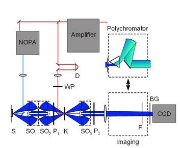
Figure 1 Schematic of the Kerr-gated optical microscope setup.
The setup allows for an instrument response function (IRF) of 85 fs when operated with a fused silica Kerr medium. A YAG Kerr medium results in an increase of the peak transmission to up to 27% while changing the IRF to around 200 fs.
ZnO nanowires were used to demonstrate the capabilities of the setup as well as the efficacy of the technique in general. ZnO has a band edge emission at 380 nm, which is well within the detection range of the new microscope and has been reported to show complex ultrafast dynamics in the UV [2]. Single ZnO nanowires grown via a vapor-liquid-solid method were investigated. Figure 2 shows luminescence images collected from a nanowire at different delay times. The evolution of the dynamics along the nanowire can be extracted along the nanowire [Fig. 2(a)].
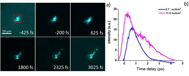
Figure 2 a) Transient image sequence of a single ZnO nanowire excited at 11.5 mJ∕cm2. (b) Transient dynamics extracted from the region on the wire demarcated by the red box in (a) for excitation fluences 9.7 mJ∕cm2 and 11.5 mJ∕cm2.
The non-linear dynamics that is observed at high excitation fluences has been attributed to the onset of amplified spontaneous emission (ASE). We have shown previously that ASE dynamics contains information about Shockley-Read, nongeminate and Auger recombination rates that give direct access to diffusion and defect limited charge carrier mobility [3]. Comparison of SEM and TEM measurements on the same wire will enable us to correlate surface morphology to changes in charge carrier mobility and can help to identify regions of high catalytic activity.
One graduate student was supported during the second year of the grant period. In addition, the project gave the opportunity for one student to perform undergraduate research. The students developed the first femtosecond time resolved UV-fluorescence microscope in this short time period. We collected valuable results that were used to revise and improve the design. Results from this project were communicated in one publication and two talks and poster presentation by the graduate student. Research activities described above have greatly impacted the professional development of the students involved due to the breadth of their experiences in the laboratory. Activities associated with this project have greatly contributed to the PI's career. He was invited to present results related to this project at the 2015 PACIFICHEM meeting. Progress in this research over the award period has led to new and broader research directions from the original proposal and the PI has been developing and submitting proposals to other funding agencies for subsequent support.
1. J. C. Blake, P. S. Eldridge, and L. Gundlach, Chem. Phys. 442, 128 (2014).
2. R. Roder, T. P. H. Sidiropoulos, C. Tessarek, S. Christiansen, R. F. Oulton, and C. Ronning, Nano Lett. 15, 4637 (2015).
3. L. Gundlach and P. Piotrowiak, J. Phys. Chem. C 113, 12162 (2009)