Reports: DNI651233-DNI6: Fundamental Antenna-Receiver Interactions in Metal-Based Light-Harvesting Nanostructures Studied by Magneto-Optical Spectroscopy
 printer friendly
Utilizing ACS funding, we have made significant research progress toward our project goals of developing a fundamental understanding of the influence of plasmon fields on molecular dynamics. Upon completion, my research group will possess the experimental and theoretical expertise to probe state-specific interactions between nanoparticle plasmon resonances and molecular electric- and magnetic-dipoles. The predictive understanding of plasmon-molecular-dipole interactions arising from this fundamental research program may result in enhanced solar photo-conversion efficiency, though light-harvesting and photocatalytic processes. In particular, our use of plasmon-enhanced magneto-optical spectroscopy can impact several areas of chemistry by providing insight into nanostructure oxidation and spin state. Progress toward these goals in three key areas are highlighted: i) implementation of continuous polarization variation (CPV) analysis to quantify nanoscale surface electric fields and magnetization components, ii) the assembly and characterization of molecular-bridged nanoparticle dimers, and iii) the development of a nonlinear optical imaging facility capable of nanometer spatial accuracy.
I. Quantification of electric- and magnetic-dipolar contributions to metal nanoparticle optical properties.
The first area of progress described in this report is the development and use of continuous polarization variation-detected second harmonic generation (CPV-SHG) to quantify the relative electric and magnetic dipolar contributions to metal nanoparticle optical properties.1-3 A significant advantage of SHG measurements over emission and linear scattering techniques is that the signal is described by the sample's nonlinear susceptibility. A general expression describing the experimentally measured SHG intensity I(2w) as a function of the polarization of the fundamental wave follows:
![]() (Eqn.1)
(Eqn.1)
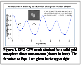 S(f) and P(f)
are the fundamental polarization states in the s and p laboratory frames; F, G,
and H are nonlinear susceptibility components. F corresponds to
electric-dipolar contributions, G to magnetic-dipolar contributions, and H to a
linear combination of the two. Therefore, SHG measurements conducted using
continuous polarization variation inform on nanoparticle surface field
symmetry.
S(f) and P(f)
are the fundamental polarization states in the s and p laboratory frames; F, G,
and H are nonlinear susceptibility components. F corresponds to
electric-dipolar contributions, G to magnetic-dipolar contributions, and H to a
linear combination of the two. Therefore, SHG measurements conducted using
continuous polarization variation inform on nanoparticle surface field
symmetry.
We extended our studies to demonstrate that the non-zero magnetic-dipole responses were the result of chiral plasmon fields activated by circularly polarized light. SHG quantification was made by circular dichroism measurements the incident fundamental light was either right or left circularly polarized and quantifying the differential SHG response. The data demonstrated large, unambiguous differences in the signal upon switching from right to left circularly polarized incident light. The extent of this chirality was determined using the SHG circular-difference ratio (SHG-CDR):
![]() (Eqn. 2)
(Eqn. 2)
The SHG-CDR can assume values between 0 and 2. In a study of several SGN dimers, we observed structure-specific non-zero CD values for many (but not all) structures. An important result from this research stage is the use of colloidal nanoparticles to amplify circularly polarized electromagnetic fields, which represents a major step toward achieving spatially resolved single-particle magneto-optical measurements. This research will enhance the understanding of electronic structure of molecules near nanoparticle surfaces and provide descriptions of interactions between plasmon modes and specific molecular states. In addition to achieving the proposed research goals, synthesis and assembly of nanoparticle dimers and development of advanced experimental facilities suited for plasmon-enhanced magneto-optical spectroscopy will allow the PI to achieve his long-term research and education objectives.
II. Molecularly-bridged SGN dimers
We published a paper on the preparation and characterization of iron porphyrin-bridged SGN dimers. SGN dimers are formed by electrostatic interactions between the porphyrin and negatively charged gold nanoparticles; the N-CH3+ groups of the iron porphyrin located in the interparticle gap between dimers interacts with citrate molecules on the SGN surface.4 Surface-enhanced Raman data indicated the porphyrin binds the metal in an edge-on geometry.4 During the next year of ACS support, we will incorporate our NLO microscope into a superconducting magnet, allowing for plasmon-enhanced magneto-optical imaging. Iron porphyrins, whose MCD spectra have been described in detail, will be used as the model system.
III. Nonlinear optical imaging with 1-nm accuracy
In continuing research, we are using plasmon
amplification to increase the spatial accuracy of nonlinear optical imaging.
These methods will be implemented to study fundamental molecular dynamics at
nanoparticle surfaces (e.g. photocatalysis). Our first paper in this area,
which described the use of plasmon amplification of nonlinear optical wave-mixing signals
to generate optical images in which the position of the scattering point source
can be determined with nanometer accuracy was published in the Journal of
Chemical Physics in 2013.5 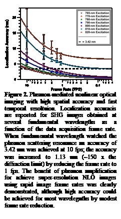 I note that this manuscript was selected
as an editor's choice article and featured on the JCP web site during the month
of July. We showed that solid gold nanosphere dimers could be used as a model
system for the nonlinear medium, which converted the Ti:sapphire fundamental to
its second harmonic frequency. Matching the fundamental wave energy to the
localized surface plasmon resonance of the electromagnetically coupled
nanospheres was critical for achieving the high localization accuracy. Our
technique, named Nonlinear Optical Localization using Electromagnetic Surface
fields (NOLES) imaging, routinely yielded nonlinear optical images with 1-nm
localization accuracy at rates ≥2 fps (Fig. 3). This high level of accuracy in pinpointing the
signal point source position exceeded that made possible using conventional
diffraction-limited far-field methods by 160×. The NOLES technique, with its high temporal resolution and
spatial accuracy that far surpass the performance typical of fluorescence-based
imaging, will be relevant for imaging dynamic chemical, biological, and
material environments.
I note that this manuscript was selected
as an editor's choice article and featured on the JCP web site during the month
of July. We showed that solid gold nanosphere dimers could be used as a model
system for the nonlinear medium, which converted the Ti:sapphire fundamental to
its second harmonic frequency. Matching the fundamental wave energy to the
localized surface plasmon resonance of the electromagnetically coupled
nanospheres was critical for achieving the high localization accuracy. Our
technique, named Nonlinear Optical Localization using Electromagnetic Surface
fields (NOLES) imaging, routinely yielded nonlinear optical images with 1-nm
localization accuracy at rates ≥2 fps (Fig. 3). This high level of accuracy in pinpointing the
signal point source position exceeded that made possible using conventional
diffraction-limited far-field methods by 160×. The NOLES technique, with its high temporal resolution and
spatial accuracy that far surpass the performance typical of fluorescence-based
imaging, will be relevant for imaging dynamic chemical, biological, and
material environments.
In addition to providing the PI with experimental infrastructure to perform fundamental plasmon-enhanced magneto-opitcal measurements on light-harvesting nanostructures, the research effort has provided training for four students (3 graduate, 1 undergraduate): Anne-Marie Dowgiallo prepared and characterized nanoparticle assemblies; Jeremy Jarrett developed SHG facilities; Patrick Herbert made significant contributions to polarization-resolved measurements; Anabel Rodriguez assisted in statistical analysis.
1. M. Chandra; A. M. Dowgiallo; K.L. Knappenberger, Jr. J.Am.Chem.Soc. 2012, 134, 4177-4180
2. M. Chandra; K. L. Knappenberger, Jr. Phys.Chem.Chem.Phys. 2013, 15, 4177-4182.
3. K. L. Knappenberger, Jr. et al. J.Phys.Chem.Lett., 2013, 4, 1109-1119.
4. L. J. Williams; A.M. Dogiallo; K. L. Knappenberger, Jr. Phys.Chem.Chem.Phys. 2013, 15, 11840-11845.
5. J. W. Jarrett; M. Chandra; K. L. Knappenberger, Jr. J.Chem.Phys. 2013, 138, 214202.