Reports: ND852265-ND8: Geophysical Monitoring of Mechanical and Chemical Alteration of Frictional Discontinuities
 printer friendly
Significant chemical and mechanical changes take place in the rock, rock joints and fluid during oil recovery procedures. The goal of this project is to show that chemical or mechanical phenomena in fractured rock, and in particular in joints, can be recognized seismically. This is achieved by performing lab-scale experiments to examine full waveforms of compressional and shear waves on fractured Indiana limestone permeated with a chemically reactive flow.
The first step of the experimental program was the characterization of Indiana limestone. Cubic specimens of the rock with 4 inches in size were subjected to uniaxial loading with concurrent measurements of transmitted compressional and shear waves. Sixteen pairs of contact compressional and shear wave transducers were used. Nine were placed at the top of the specimens as receivers and nine at the bottom as sources. The other seven pairs were attached to the sides. Honey was used as the couplant between the rock and each transducer. Tests on aluminum, used as a standard, showed that the honey couplant stabilized after 200 minutes when placed in a loading frame under a constant stress of 15.5 MPa. At the end of this time period, the peak to peak amplitudes of the waveforms became constant for all transducers. The same test on Indiana limestone showed the same results. The repeatability of the tests was assessed by conducting each test three times. As a result, the experimental errors were assessed as follows: due to spatial variability, 0% to 3% in wave velocity, and 0% to 4% in peak-to-peak amplitude for Indiana limestone. For aluminum, the errors were much smaller, of about 0% to 2%.
Material variability, that is the difference in response from specimen to specimen, was evaluated by conducting additional tests. In the experiments, a stress of 15.5 MPa was applied in ten steps and then decreased to 0 in another ten steps. In each step, seismic response was monitored. Tests were also conducted on the same specimens but rotating the load and position of the seismic array, i.e. the load was applied at different faces while the seismic transducers were attached to also different faces than in previous tests. The variation of peak-to-peak amplitude and velocity with loading ranged from 8% to 30% and 0% to 2%, respectively, for Indiana limestone. For aluminum, the variation was 0%. Each loading cycle was repeated three times with results from all cycles indicating the same trend.
Fractures were artificially induced to intact blocks of limestone using a modified Brazilian loading where cubic specimens of the rock (4x4x4 inches) were loaded in compression from opposite faces. To investigate the suitability of the technique, a series of gypsum cubes, with the same dimensions as the Indiana limestone specimens, were made first. This was accomplished by mixing gypsum (Hydrocal B11 from U.S. Gypsum Company), diatomaceous earth and water. The mass proportions in the mix were water/gypsum = 0.6 and water/diatomaceous earth = 35. The gypsum cubes were placed in a load frame, with aluminum rods between the loading platens and the top and bottom faces of the cubes to induce a stress concentration at the point of application of the load. The load was applied at a slow rate until failure. Different loading rates and rod diameters were used, with the best results obtained at 25 lbs/sec and 13 mm rods. This technique was then used to fracture the Indiana limestone cubes with excellent results. A laser profilometer was used to measure the surface roughness by taking measurements at 0.25 mm increments in two orthogonal directions.
A set of fluid invasion tests was conducted on intact and fractured column specimens of Indiana limestone. The dimensions of the column specimens were 2 × 2 × 4 inches. Water was injected through the bottom of the intact specimen and through one of the sides of the fracture, on the fractured specimen. As the water flowed through the intact limestone and/or through the fracture, transmitted compressional and shear waves across the intact specimen and across the fracture were monitored. Figure 1 shows the normalized peak-to-peak amplitudes of compressional waves (left) and shear waves (right) on the fractured specimen; similar observations were made on the intact specimen. The magnitude of the amplitude of each transducer is normalized with respect to its value at the beginning of the test (i.e. when the material and/or fracture were dry). The figure shows a first segment, from about time zero to about time 100 minutes depending on the transducer, of constant amplitude. Then there is an abrupt decrease of amplitude to about 50% to 60% of its original value, followed by a plateau at larger times. Our interpretation is that the dramatic decrease of amplitude is associated with the fluid front arriving at the imaginary line connecting the corresponding transducer pair. Further analysis of the data (not shown well in Figure 1) clearly indicates that detection of the front (drop of amplitude) happens first at those transducers closer to the inlet location and then later at transducer pairs with increasing distance from the inlet. This has resulted in a linear correlation (not shown in this report) between time and location of the transducer pairs. This has allowed the computation of the water invasion velocity, which has resulted in velocities of 0.005 cm/sec. through the intact rock (at 10 KPa of water pressure) and 0.003 cm/sec. through the fracture (at 0.1 Pa). These tests have provided two important observations: (1) seismic methods are effective in detecting the invasion front of a fluid through a discontinuity and/or through the intact material; and (2) sealing of the intact material in tests with flow through a fracture is imperative, as the flow velocity inside the rock is as high as through the fracture. Work is currently underway to develop a methodology to effectively isolate the fracture from the rest of the specimen to conduct fluid tests through the fracture at higher heads.
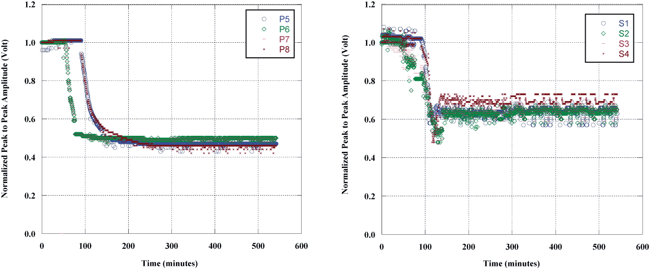
Figure 1. Peak-to-peak amplitude during fluid invasion. P- (left) and S- (right) wave transducers.