Reports: ND555120-ND5: A Microrheological Study of Interfacial Nanoparticle Layers: Understanding Glassy Rheology, Aging and Stability
 printer friendly
Overview
The goal of this project is to understand the interfacial rheology of dense collections of amphiphilic nanoparticles on oil water interfaces, informed by the hypothesis that they may resemble soft glasses, such as foams and slurries. An earlier study probed Gibbs layers formed from these suspensions in a pendant drop apparatus, determining the equilibrium pressure-density isotherm and interfacial particle pair potential. The use of the pendant drop method, however, precluded the measurement of interfacial rheology. The current study seeks to probe this system using state of the art interfacial microrheology which will require small probes to provide the needed sensitivity.
In the first funding period, with postdoc Dr. Mehdi Molaei, we developed a compact, 3-d printed device with many of the functions of a Langmuir trough, but well suited to high performance microscopy, and developed a laser-illuminated dark-field microscope for imaging and tracking gold-nanorods. In the second funding period we have performed two studies with these instruments, the first advancing the state of the art by using gold nanorods as tracers for rotational microrheology and the second performing simultaneous interfacial tensiometry and microrheology on a Gibbs layer of b-lactoglobulin on an air-water interface.
Rotational microrheology with gold nanorods
Passive microrheology, developed more than 20 years ago, typically quantifies the Brownian motion of microparticles embedded in a viscoelastic fluid with a mean-squared displacement and then converts it numerically into a frequency-dependent shear modulus. A major limitation of microrheology is that for stiff elastic materials, the amplitude of the motion can become very small. We strove to develop the 'ultimate' microrheology method, by tracking the rotational Brownian motion of a convenient tracer, a gold nanorod (GNR), using our recently developed dark-field microscope.
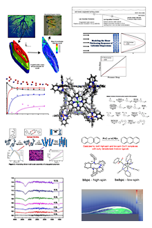
This project required multiple innovations. First we modified our dark-field microscope to separate the light scattered from the nanorods into two orthogonal polarizations, forming two separate images side by side on the same detector of a high-speed CMOS camera. We derived a quantitative nano-optical model that related the GNR's brightness in these two images to its orientation in three-dimensional space; the results were accurate to better than 1°. Typical results for 20nm diameter and 100nm long GNRs in glycerol shown in Figure 1. This method was then used to determine the mean-squared angular displacement of the Brownian tumbling of single nanorods. Because our tracking method requires inverting a periodic mapping function, however, these orientations are mapped into a single octant of the unit sphere, and must be converted into an unconstrained form suitable for use in the rotational Stokes-Einstein relation.
In the end, we demonstrated this method with the same GNR tracers in viscoelastic polymer solutions, and obtained results that agreed with macroscopic rheology results. These measurements advanced the speed and stiffness limits for rotational microrheology by orders of magnitude relative to earlier reports, while measuring bulk shear moduli on a single attoliter (10-18l) volume. These experiments are described in a manuscript currently under review.
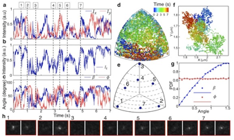
| Figure 1: (a) Measured scattering signal from x (blue) and y (red) channels of a GNR embedded in glycerol. (b) The total intensity of the GNR signal. (c) Resolved polar angles (blue) and azimuthal angles(red) of the GNR. (d) 3D orientation of the GNR, location of ,u.(t), colored by time. (f) 2D trajectory of the GNR colored by time showing a translational Brownian motion. (g) Measured probability distribution of polar angles (blue circles) and azimuthal angles (red squares) from 105 image pairs showing good agreement with expected distribution for a GNR randomly exploring a unit octant, solid lines. (h) Sample images of the GNR at different orientations, (shown in e with blue circles) at selected times (shown by dashed line in a-c).
|
Interfacial tensiometry and microrheology of adsorbed protein films
A convenient and interesting model system for studying interfacial rheology consists of a protein spontaneously adsorbing onto a water-air interface. It is expected that most proteins upon adsorption will undergo denaturation, making them likely to aggregate laterally with other adsorbed protein molecules. Literature findings on such systems, e.g. the milk protein b-lactoglobulin, find that at long times an elastic and plastic interfacial film forms, whose elasticity appears suddenly in a manner expected for a percolation process. Interfacial rheometry prior to that percolation point gives seemingly contradictory results, which have been interpreted variously in the literature. We thought that this system would allow an excellent first demonstration of the capabilities of our 3-d printed trough (3DPT), which allows simultaneous tensiometry, microrheology and perturbation of surface density.
The first question we sought to answer was the nature of the interfacial protein layer prior to the moment of percolation. Typical microrheology data are presented in Figure 2A, which show diffusive behavior at short times, with diffusivities that drop slightly compared to the clean interface value, and which then drop abruptly to a low value and a predominantly elastic behavior at t = 660 second. Based upon this analysis alone, we would conclude that the interface is viscous, perhaps with individual adsorbed protein, or perhaps disconnected clusters of aggregated protein (pre-gel). Tensiometry (not shown) reveals a surface tension comparable to a clean interface, consistent with the low surface pressure expected in the pre-gel state. Remarkably, perturbing the interfacial area reveals a significant and elastic extensional modulus, Figure 2B. How can the interface be both elastic (under extension) and viscous (under microrheology)? The idea that the tracers are occupying holes in the film seems unlikely given that they can move many microns at long times, comparable to the their mean spacing, rather than displaying cage diffusion. We are currently performing two-point microrheology measurements to understand this apparent conundrum.
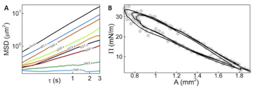
| Figure 2: A Shows typical mean-squared displacements of 1 µm microspheres in an air-water interface, as b-lactoglobulin diffuses to the surface, showing the onset of elasticity ~660 seconds. B Shows early-time surface pressure as the (initially 1.8 mm2) interface is alternately compressed and expanded, showing a fully reversible and not quite purely elastic extensional modulus for the interfacial layer.
|