Reports: DNI655184-DNI6: Visualizing Molecular Organization and Energy Transport Dynamics at Organic Surfaces and Heterojunctions with Interface Specific Femtosecond Spectroscopy

Sean T. Roberts, PhD, University of Texas, Austin

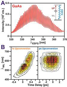
Organic semiconductors (OSCs) are a unique class of petroleum-derived materials that blend the processing advantages of plastics with the electrical properties of semiconductors. In contrast to inorganic semiconductors such as silicon, OSCs can be readily processed from solution into highly-absorbing, thin, conductive films, making OSCs attractive for a wide range of optoelectronic applications. In many of these applications, the transfer of charge to and from OSCs is fundamental to device operation. Our goal is to develop interface selective spectroscopies that can investigate OSC interfaces and establish how the molecular organization of these regions controls their ability transfer charge. The suite of techniques we have worked to develop utilize electronic sum frequency generation (ESFG) to achieve interfacial selectivity. In an ESFG measurement, two pulses of light, a white light (WL) continuum and a narrowband upconversion pulse illuminate a sample, producing a new light pulse at their sum frequency (Figure 1A inset). Resonance between frequency components of the WL and electronic transitions within a sample strongly enhances this mixing, allowing ESFG to serve as a proxy for the sample’s electronic absorption spectrum. Importantly, due to the cancellation of forward- and back-propagating fields, ESFG is preferentially created by regions of a sample that lack inversion symmetry. As OSC interfaces naturally lack such symmetry, ESFG can selective examine the structure and dynamics of these critical regions. Over the last year, we have achieved two key milestones described below:

Construction of a Spectrometer for Rapid ESFG Acquisition

One of our goals is to develop ESFG into a time-resolved probe that can follow photoinduced electron transfer at OSC interfaces. This requires differencing ESFG spectra measured before and after OSC photoexcitation for multiple time delays between an excitation pulse and an ESFG probe. Unfortunately, measuring an ESFG spectrum using our initially reported spectrometer design takes ~1 hour due to both the low photon generation rate of ESFG signals and the need to scan multiple time delays between the WL continuum and upconversion beam due to spectral chirp in the WL. Over the past year, we have implemented two key upgrades that rapidly speed data acquisition. First, we have employed heterodyne detection, which uses a reference field to amplify signals produced by sample OSC films (Figure 1A). Details regarding this collection scheme were published in J. Phys. Chem. C in August. Secondly, we have exchanged the femtosecond upconversion pulse in our initial spectrometer design with a picosecond pulse produced by an optical parametric amplifier that can upconvert the full bandwidth of the WL continuum without the need to scan the time delay between the pulses (Figure 1B). Together, these two upgrades have decreased ESFG data acquisition times to ~2 minutes. We believe further gains are possible via improvements to mirrors and optics to reduce reflection and transmission losses. We are now working to integrate a photoexcitation source in-line with our ESFG spectrometer to investigate electron transfer between perylenediimide films and silicon.

Figure 1: (A) ESFG spectrum of GaAs(110) measured with heterodyne detection. Fringes result from interference between the GaAs signal and the reference field that amplifies it. (inset) WL and upconversion fields (Up) mix in GaAs to produce ESFG. (B) ESFG signal of z-cut quartz as a function of the time delay between the WL and Up fields. While the ESFG spectrum changes with this delay when using a ~100 fs Up field, a 3 ps Up pulse removes the need to scan this delay.

Examining the Electronic Structure of Perylenediimide Interfaces

Perylenediimides (PDIs) are dye molecules that display semiconducting behavior when assembled into films. Due to their high photostability, molar extinction, and electron accepting character, PDIs are common components in OSC devices. Recently, these materials have attracted interest due to their ability to undergo singlet fission (SF), a process wherein a photoexcited spin-singlet exciton uses its energy to create two spin-triplet excitons on neighboring molecules. As SF effectively uses a single photon to excite two electrons, SF can lower thermalization losses in both photovoltaic and photocatalytic technologies. We have been working to investigate schemes to extract triplet excitons from PDI films; in particular, by transferring these excitons into inorganic semiconductors such as silicon. Understanding how the electronic structure of PDI films is modified at PDI:silicon interfaces is key to this approach’s success. Over the past year, we have used ESFG to study how processing conditions and the side chains of different PDIs alter their packing structure at SiO2 interfaces in preparation to examine PDI:silicon surfaces.

Figure 2B compares the electronic absorption spectrum of N-N'-Dioctyl-3,4,9,10-perylenedicarboximide (C8-PDI) against its ESFG spectrum and shows the ESFG spectrum is shifted to lower frequency. We have observed similar behavior in other PDIs, notably N-N'-Dimethyl-3,4,9,10-perylenedicarboximide (C1-PDI), which we described in the J. Phys. Chem. C article where we detailed our HD-ESFG setup. We can rationalize this shift by noting PDI films are subject to compressive strain at a SiO2 interface, which can shift the intermolecular spacing between adjacent PDIs. The broad bulk absorption spectrum seen in Figure 2B results from electronic coupling between adjacent PDIs that cause their frontier molecular orbitals to extend over multiple molecules. Slipping neighboring PDIs with respect to one another due to interfacial strain will alter this coupling, leading to changes in their electronic structure and a shifted ESFG spectrum. Plotted alongside the experimental traces are calculations based on a Holstein Hamiltonian that predict how the optical spectrum of C8-PDI is expected to change when molecules are slipped by 0.5 Å along their short axis from the structure they adopt in bulk films. These calculations capture the observed spectral changes exceptionally well, and suggest C8-PDI molecules change their intermolecular packing at buried interfaces. We are now extending these measurements to PDI films on methyl-terminated Si(111). This work will continue with support from an NSF CAREER award, which was earned on the basis of preliminary results obtained via our DNI grant.

Figure 2: (A) PDIs form slip-stacked columns in the thin films. (B) Absorption (blue) and ESFG spectra (red) of a C8-PDI film plotted alongside calculated spectra (dashed black).