Reports: DNI853833-DNI8: Organic Carbon Preservation in Deep-Time: Exceptional Glimpses from Exceptionally-Preserved Fossils and Laboratory-Based Decay Experimentation
James D. Schiffbauer, PhD, University of Missouri
The primary focus of this PRF DNI research was to examine first-order controls of Burgess Shale-type fossil preservation. To do so, my laboratory group has employed two complementary approaches: (1) analysis of Burgess Shale-type fossil materials, and (2) controlled laboratory- and field-based decay experimentation. Burgess Shale-type preservation, named for the famed Cambrian fossil locality, reveals exceptional glimpses of the aftermath of the first major diversification of animals—the Cambrian Explosion—captured largely as two-dimensional carbonaceous compressions of labile tissues, though commonly associated with accessory mineralization. In the broadest sense, generally similar in focus to understanding the formation of petroleum or other carbon-bearing resources, the overarching goal of this research effort was to evaluate the diagenetic, biological, and paleoenvironmental factors responsible for generating carbonaceous compression fossils.
1. Fossil materials—
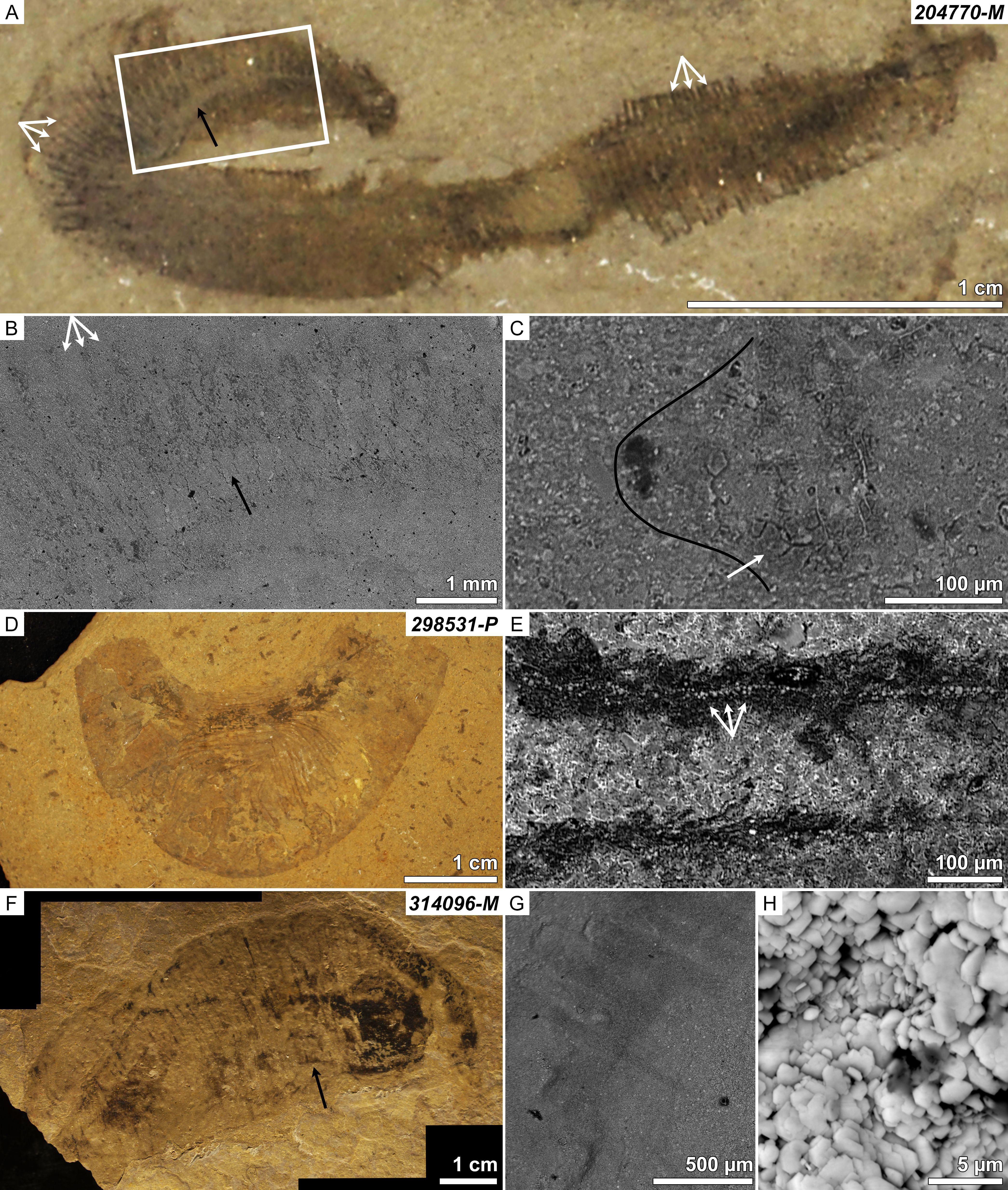
Over the last grant period, which was a no-cost extension to finalize data collection with remaining funds, the PRF DNI funding was utilized to complete scanning electron microscopy-based microchemical analyses of fossil and decay experiment materials. In addition to two previous publications in the Journal of Paleontology, we have published a third paper in the September 2017 issue of Palaios, led by a PhD student author, reporting comparative analyses of Burgess Shale-type vermiform (worm-like) fossils from the western US (see Fig. 1, Broce and Schiffbauer, 2017). As with our previous publications, insights gained from this work have contributed greatly to the design of laboratory- and field-based decay experimentation, as briefly described below.
2. Laboratory- and field-based decay experimentation—
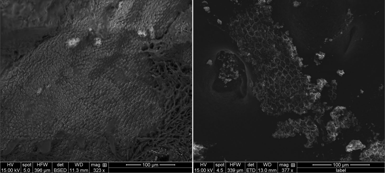
This grant has additionally supported the operation and maintenance of an anoxic glove-box in my decay laboratory, in which modern analogs of the analyzed fossil materials have undergone decay experiments to observe early stages of “preservation” on laboratory timescales (see Fig. 2, repeated from the 2015–2016 annual report). Although impossible to replicate geologic time, this protocol allows for actualistic observation of the pace of decay under conditions designed to mimic the Cambrian seafloor where their fossil counterparts were preserved. At present, we have completed decay experimentation on waxworms (caterpillar larvae of the wax moth) and shrimp, which should result in a publishable report, and are additionally finalizing manuscript preparation reporting the results of our field-based decay experimentation of crabs, conducted in a natural coastal environment at the Gerace Research Center, San Salvador, Bahamas.
3. Summary—
This serves as the final report for this PRF DNI funding, but several important notes should be added. In summation, this PRF DNI funding will have: (1) resulted in the production of at least 5 published manuscripts; (2) supported several (n=3) graduate students both at the MS and PhD levels as well as one undergraduate researcher; and (3) resulted in the dissemination of our results widely at national meetings through student-given presentations (n>20). Perhaps most importantly, however, this PRF DNI funding has built firm foundations that have helped in procuring long-term research funding: I was recently awarded an NSF CAREER Award, which began in August 2017 and will directly build upon the initial studies conducted here.
4. Reference cited—
Broce, J.S., and Schiffbauer, J.D. 2017. Taphonomic analysis of Cambrian vermiform fossils of Utah and Nevada, and implications for the chemistry of Burgess Shale-type preservation. Palaios 32: 600–619.
5. Figures and captions—
Figure 1: Kerogenized vermiform specimens, all of which belong to the genus Ottoia, from the Cambrian western US collections at the University of Kansas Biodiversity Institute (KUMIP). (A–C) are KUMIP 204770-M; (D–E) are KUMIP 298531-P; and F–H are KUMIP 314096-M. (A) Optical photomicrograph of 204770-M; (B) BSE image [from rectangle area in (A)] of annulations (white arrows) and gut (black arrow); (C) kerogen patches and clay textures inside vs. outside of fossil material. Note cracked texture of kerogen patches due to volumetric reduction (white arrow), and smoother, finer-grained nature of clays associated with fossil material (right) as compared to those of the host rock (left). Black line corresponds to body wall. (D) Optical photomicrograph of 298531-P; (E) thin line of framboidal pyrite (white arrows) within bands of inert kerogen (darker surrounding material). (F) Optical photomicrograph of 314096-M; (G) bands of different clay textures preserving annuli [from black arrow region in (F)], note differing greyscale; (F) detail of bladed barite patches.
Figure 2: Hexagonal surface ornamentation on waxworm (left; backscattered electron image in low vacuum mode), and hexagonal molds in iron sulfide minerals (bright hexagonal shapes) from recovered from decay environment (right; secondary electron image in low vacuum mode).

