Reports: ND1055283-ND10: Micro/Nanoscopic Investigation of ZnO towards the Development of Next-Generation Separation Platforms
 printer friendly
The overall goal of the project is i) to generate well-defined zinc oxide (ZnO) nano and microstructures in the forms of 2D (thin film), 1D (nanorods, microrods), and 0D (nanoparticles, microparticles) and ii) to examine their crystalline facet-specific chemical reactivity for the design of better separation platforms.
After establishing reliable synthetic routes to produce various forms of ZnO materials, we have characterized the as-synthesized (or after purification) materials by spectroscopic and imaging tools such as SEM, AFM, pXRD, EDAX, ATR/FTIR, UV-Vis, and Raman. Among the various ZnO forms produced, the NR ensemble samples were further exploited as an active channel in a photodetector device operating in the visible wavelength range. The ZnO NR-based devices demonstrated extremely high electrical responses upon light illumination. The outcomes of this endeavor have been reported in Nanotech. 28, 145203, (2017) and a provisional patent application was filed.
D. S. Choi, M. Hansen, E. V. Keuren, and J. Hahm, Highly photoresponsive, ZnO nanorod-based photodetector for operation in the visible spectral range, Nanotechnology, 28, 145203 (2017).
J. Hahm, D. S. Choi, M. Hansen, E. V. Keuren, ZnO Photodetector, U.S. Provisional Patent Application No. 62/426,055 (2016).
We have also begun to examine the potentially different degrees of chemical reactivity depending on the crystalline facets present within individual ZnO nano- and micro-materials. In addition to the direct imaging methods of AFM and SEM which can provide information on the morphological changes of the crystal surface, an optical scattering-based method providing a sub-crystal-level spatial resolution was used to determine position- and crystalline facet-dependent optical responses. An example based on elastic scattering is shown in the figure below, which was obtained for the case of Mie scattering from a ZnO NR.
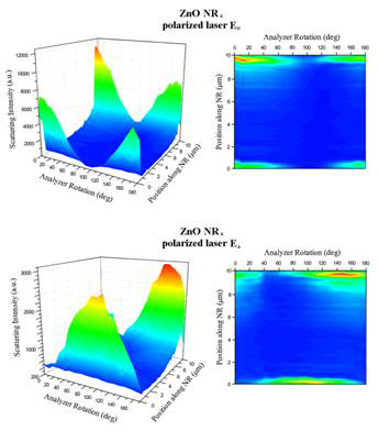
Figure III. Scattering of a single ZnO NR measured by using two polarization directions of an incoming laser (E║ and E┴) on a NR oriented along the x-axis (ZnO NR┴). Scattering intensity was measured with respect to the position along the length of the 1D nanomaterial as well as the analyzer angle.
An inelastic scattering technique of Raman has been increasingly exploited as a noninvasive and sensitive analytical tool to investigate material properties pertinent to separation and sorption. We have also employed Raman to examine as grown ZnO nano- and micro-structures as well as those reacted with various sulfur sources in gas and aqueous phases. Firstly, we carefully examined unreacted forms of individual ZnO structures in order to discern any material position-dependent signal differences in Raman intensity. This position-dependent signal difference becomes important for the ZnO NR crystals since their high shape anisotropy (length to width) can lead to unusual scattering behaviors at the two ends of the NRs relative to the main body. Characterizing such position-dependent behaviors will become crucial for the accurate determination and interpretation of the facet reactivity differences measured from sulfur-reacted ZnO crystals. Hence, for the first time, we performed a systematic investigation to understand the Raman outcomes of the highly anisotropic ZnO NRs versus isotropic ZnO NPs/MPs at the individual nanomaterial level. An example of our major findings can be seen from the Raman mapping data presented below for ZnO NRs and ZnO MPs. The detailed results from this study can be found in Nanoscale, 9, 8470-8480, (2017).
M. Hansen, J. Truong, T. Xie, and J. Hahm, Spatially distinct Raman scattering characteristics of individual ZnO nanorods under controlled polarization: Intense end scattering from forbidden modes, Nanoscale, 9, 8470-8480 (2017).
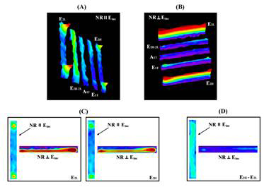
Figure IV. Raman intensity maps of the five ZnO phonon modes identified from a ZnO NR are displayed combinedly for the different light-matter interaction geometries. The data show strongly NR position-dependent Raman scattering profiles for the minor phonon modes per given orientation, whereas the major modes for each orientation display continuous and persistent Raman intensities distributed all along the entire NR length.
In this work, we quantified Raman signals from the five key ZnO phonon modes of E2L, E2H-2L, A1T, E1T, and E2H, and revealed the NR position-dependent Raman scattering characteristics of the phonon modes per given light-matter interaction geometry. We then presented Raman intensity maps and elucidate Raman behaviors consistent and incongruous with Raman selection rules. In particular, we have identified an intriguing Raman scattering phenomenon from the forbidden modes, distinctively occurring at the two NR ends. Their unexpectedly strong and localized scattering signals at the NR termini were contrasted by the scattering behaviors from the rest of the NR positions agreeing with the selection predictions. Subsequently, by carrying out control measurements on isotropic ZnO MPs, we ascertained that the unique NR position-specific Raman responses observed on ZnO NRs originated from their high shape anisotropy.
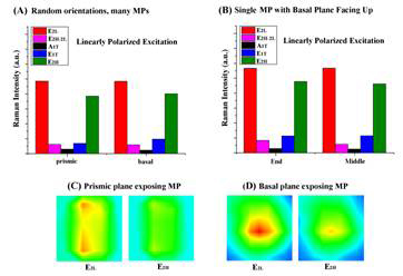
Figure V. Raman data collected from isotropic ZnO MPs are summarized. The same trend in the Raman mode intensities were observed regardless of the different positions on each facet examined. In all cases, the strongest signals were produced by the E2L and E2H modes.
We are now in the final stage of performing combined measurements on sulfur-reacted samples of ZnO NRs and NPs/MPs in order to determine the chemical reactivity associated with various crystal facets. Imaging, spectroscopic, scattering, as well as optical measurement techniques are being used for this investigation which include SEM, EDAX, TEM, AFM, XRD, UV-Vis, ATR/FTIR, PL, and Raman. We will report the new findings of these studies when the study is completed in the next fiscal year.