Reports: ND1053615-ND10: Atom Probe Tomography of Kerogen and the Kerogen-Mineral Interface
Derk Joester, PhD, Northwestern University
Executive Summary. The most abundant form of organic carbon on Earth, kerogen, is a mixture of insoluble macromolecules dispersed in sedimentary rocks. Remarkably stable against environmental and biological degradation, it is not only crucial as a source of oil and gas, but also a source of information on the paleo-environment. However, our understanding of the structural features of the macromolecules we refer to as kerogen is rather limited. We proposed to use atom probe tomography (APT) to investigate kerogen structure. During our investigation, we quickly realized that in order to make progress, a better understanding of the actual spatial resolution of APT for was required. We decided to follow an opportunity to do so in two systems, magnetite (Fe3O4) and apatite (Ca5(PO4)3(OH)). A systematic investigation of crystallographic poles in 3D reconstruction of geologic magnetite with exsolution features allowed us to identify features of the field-evaporation process that lead to a specific artifact in spinel-type materials (2 publications in preparation). In parallel, we used dental enamel, a biological, nanostructured apatite with a well-known structure but poorly understood interfacial chemistry in order to calibrate APT of phosphates and understand better the behavior of organics at surfaces. This lead to the discovery of an amorphous intergranular phase in dental enamel (2 papers published1-2, 2 in preparation).
Project I. Validating 3D Reconstructions of Geological Samples
APT is unrivaled in its ability to quantitatively image the distribution of atoms in a sample. The ability to detect, identify, and quantify crystallographic order in APT reconstructions is the most important internal reference for accuracy of reconstruction3-8 and determination of resolution5-6,9. This is of particular relevance for complex oxides, phosphates, and sulfides, that only recently have moved into the scope of APT10-12. When we started working on shales as we had originally proposed, we quickly realized that we were lacking information necessary to reconstruct samples with confidence. We therefore backtracked to a system we understand well, magnetite (Fe3O4, inverse spinel structure), where around the same time, in collaboration with Dr. Krishna Rajan (ISU) we discovered crystallographic poles, i.e. evidence for crystallographic ordering that could be used constrain reconstructions.
One pole, however, insufficiently constrains the reconstruction. We attacked this problem by a systematic analysis of oriented single crystals of geological magnetite, combined with advanced spatial distribution map (SDM)13 techniques to identify the orientation of poles. This allowed us to determine reconstruction parameters that rendered the angle between the poles correctly and unambiguously (manuscript in preparation).
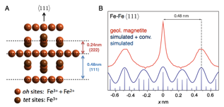
Fig. 1. A Spinel structure as seen parallel to the {111} planes, with oxyanion sublattice omitted for clarity. B Experimental and simulated Fe-Fe SDMs in the [111] direction. Comparison of the simulated SDM convoluted with the experimental one reveals that the experimental spacing is twice the expected value. Figure from MS Thesis of Michael Cohen.
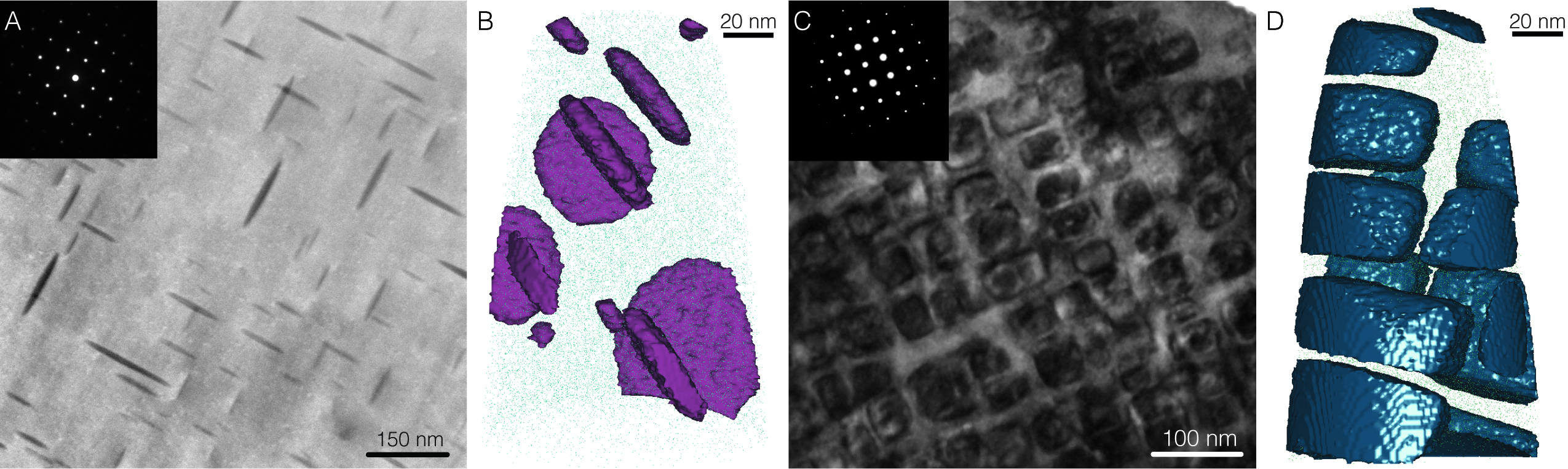
However, the SDM reported a lattice spacing precisely double the expected value (Figure 1). To better understand why this is the case, we analyzed two geological magnetites with nanoscale, coherent precipitates. In LP204-1, disk-shaped precipitates are present on the {100} planes, in a titanomagnetite sample there are cuboidal magnetite precipitates in a Ti-rich ulvšspinel matrix (Figure 2). We used geometric information gleaned from correlative STEM-HAADF to constrain APT reconstructions, and thus independently confirmed that the planar spacing of [111] poles is twice the expected value. In collaboration with Dr. Dan Schreiber (PNNL), we confirmed that this artifact also affects other spinels. An analysis of trevorite (NiFe2O4) indicated that the most likely mechanism is preferential evaporation of two consecutive layers at once. This is a significant finding in that it highlights the importance of using correlative imaging and internal references such as crystallographic poles and coherent precipitates in the reconstruction of geologic samples (manuscript in preparation).
Fig. 2. STEM-ADF images (A,C) and APT 3D reconstructions (B,D) of disk-shaped, Mg/Al/Mn rich precipitates in LP204-1 magnetite (A,B); Magnetite blocks in a Ti-rich ulvšspinel matrix (C,D). Figure adapted from MS Thesis of Michael J. Cohen.
Project II. Mineral-Organic Interfaces and Interphases in Apatites
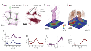
One of our original tasks was to image the kerogen-mineral interface. Given our difficulties in reconstructing clays, we decided to attempt mapping organics on the surface of apatite crystals in tooth enamel. During the formation of dental enamel, nanowires of hydroxylapatite grow in an organic gel. Proteins in the gel are degraded and were thought to be incorporated as defects at grain boundaries. As in the case of kerogen-clay interactions, it had been proposed that a monolayer of organic matter surrounds individual apatite nanowires. Similarly, Mg2+ and CO32- ions were thought to segregate to the same grain boundaries. However, determining the distribution of organic matter and impurity ions in enamel remained a very challenging problem. Unlike the shales, however, the length scale of interfacial features in enamel is such that correlative imaging by APT and electron microscopy techniques allowed us to constrain reconstructions with good accuracy (Figure 3).
Fig. 3. APT 3D reconstructions of grain boundaries (A-D) and interphases (E-H) in rat enamel (A, B, E, F) and pigmented beaver enamel (C,D,G,H). Figure adapted from Gordon et al, Science 2015.2
Our investigation into the grain boundary chemistry of dental enamel revealed a great deal of new information that went far beyond our original vision. We discovered that in rodent enamel, Mg2+ is predominantly located in intergranular precipitates of Mg-substituted amorphous calcium phosphate.2 Similarly, residual organic matter and carbonate ion is enriched in this amorphous intergranular phase.1 Remarkably, in the pigmented enamel of certain rodents, a mixture of ferrihydrite and amorphous iron-calcium phosphate replaces the much more soluble Mg-ACP. This substitution renders pigmented enamel both mechanically harder and significantly more resistant to acid attack. The existence of (metastable) amorphous intergranular phases in apatites had never before been observed. Our discovery will help understand the formation of carious lesions, aid in developing early detection schemes and enable development of "post-fluoride" dental care products and was published in Science and in Frontiers in Physiology.1-2 Several more manuscripts on more technical aspects of APT of apatites are currently in preparation.
 printer friendly