Reports: DNI651233-DNI6: Fundamental Antenna-Receiver Interactions in Metal-Based Light-Harvesting Nanostructures Studied by Magneto-Optical Spectroscopy
 printer friendly
I. Quantification of electric- and magnetic-dipolar contributions to metal nanoparticle optical properties.
Figure 1. Left: comparison of SHG image contrast obtained from a single chiral plasmonic nanostructure excited using left (top) and right (bottom) circularly polarized light. Right: normalized SHG plotted versus complete polarization variation along with the fit to Equation 1 given in the text. The fit yields large relative values for the fit parameter, G, reflecting magnetic dipolar contributions to SHG.
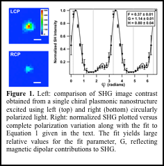
The first area of progress described in this report is the development and use of continuous polarization variation-detected second harmonic generation (CPV-SHG) to quantify the relative electric and magnetic dipolar contributions to metal nanoparticle optical properties.1-3,5,6 A significant advantage of SHG measurements over emission and linear scattering techniques is that the signal is described by the sample's nonlinear susceptibility. A general expression describing the experimentally measured SHG intensity I(2ω) as a function of the polarization of the fundamental wave follows:
![]() (Eqn. 1)
(Eqn. 1)
S(φ) and P(φ) are the fundamental polarization states in the s and p laboratory frames; F, G, and H are nonlinear susceptibility components. F corresponds to electric-dipolar contributions, G to magnetic-dipolar contributions, and H to a linear combination of the two. Therefore, SHG measurements conducted using continuous polarization variation inform on nanoparticle surface field symmetry.
An important result from this research stage is the use of chiral, colloidal nanoparticle assemblies (Figure 1) to amplify circularly polarized electromagnetic fields. This represents a major step toward achieving spatially resolved single-particle magneto-optical measurements. This research will enhance understanding of electronic structure of molecules near nanoparticle surfaces and provide descriptions of interactions between plasmon modes and specific molecular states. In addition to achieving the proposed research goals, development of advanced experimental facilities will allow the PI to achieve his long-term research and education objectives.
II. Nonlinear optical imaging with nanometer precision
Figure 2. (a): Statistical determination of the second harmonic hotspot for a nanoparticle dimer. Localization analysis with 1-σ (cyan) and 2-σ (magenta) confidence. (b) Summary of obtainable nonlinear optical localization precision obtained for several frame rates and laser excitation wavelengths. The data indicate NLO hotspots can be determined with 1-nm spatial precision with rapid image frame rates (2 frames per second).
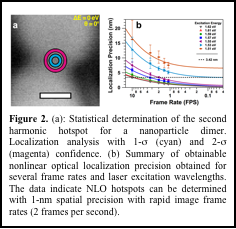
We are using plasmon amplification to increase the spatial accuracy of nonlinear optical imaging. These methods will be implemented to study fundamental molecular dynamics at nanoparticle surfaces (e.g. photocatalysis). Our first paper in this area, which described the use of plasmon amplification of nonlinear optical wave-mixing signals to generate optical images in which the position of the scattering point source can be determined with nanometer accuracy was published in the Journal of Chemical Physics in 2013. This manuscript was selected as an editor's choice article for the year 2013. We showed that solid gold nanosphere dimers could be used as a model system for the nonlinear medium, which converted the Ti:sapphire fundamental to its second harmonic frequency. Matching the fundamental wave energy to the localized surface plasmon resonance of the electromagnetically coupled nanospheres was critical for achieving the high localization accuracy. Our technique, named Nonlinear Optical Localization using Electromagnetic Surface fields (NOLES) imaging, routinely yielded nonlinear optical images with 1-nm localization accuracy at rates 2 fps (Fig. 2). This high level of accuracy in pinpointing the signal point source position exceeded that made possible using conventional diffraction-limited far-field methods by 160×. The NOLES technique, with its high spatial accuracy and data acquisition rates that far surpass the performance typical of fluorescence-based imaging, will be relevant for imaging dynamic chemical, biological, and material environments.
III. Femtosecond time-resolved nonlinear optical imaging with nanometer precision
Figure 3. SHG-detected interferogram of plasmon resonant excitation of a single nanostructure imaged with 1-nm spatial precision as shown in Figure 2.
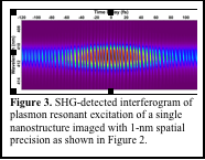 Because of the unique optical imaging capabilities developed in our lab as part of the ACS-PRF project, we have established several important collaborations. An especially fruitful one involves the Vaia (AFRL) and Nealey (U.Chicago) research groups. The objective of our work is to use femtosecond time-resolved imaging with nanometer spatial precision to monitor and control energy transfer in rationally designed nanostructures. This research effort is a natural extension of our ACS-PRF-supported program, and we expect our advances to result in predictive control over nanoscale energy flow. In order to carry out nonlinear optical imaging with both high spatial precision and femtosecond temporal resolution, we employ a collinear phase-locked pair of ultra-short laser pulses. This method has permitted us to image plasmon fields confined to a few-nanometer region with 15 femtosecond temporal resolution. A sample interferogramis given in Figure 3.
In addition to providing the PI with experimental infrastructure to perform fundamental plasmon-enhanced measurements on light-harvesting nanostructures, the research effort has provided training for several graduate and undergraduate students.