Reports: DNI753116-DNI7: Atomic-Scale Visualization of Paraffin Melting and Crystallization with Ultrafast Transmission Electron Microscopy
David J. Flannigan, PhD, University of Minnesota
This goal of this project is to establish the importance
of direct visualization – with atomic-scale spatial and picosecond to millisecond
temporal resolutions – of n-paraffin melting, nucleation, and crystal
growth with and without polymer additives such as ethylene-vinyl acetate (EVA),
a commonly-employed pour-point depressant for mitigation of waxy build-up in
crude oil infrastructure. In general, polymer melting and crystallization
involves discrete but connected processes occurring over large spatial and
temporal ranges; atomic-scale structural rearrangements such as bond rotations
occur in picoseconds, while microscale lamella formation may take milliseconds
or longer. By mapping the structural dynamics across these spatiotemporal
ranges under conditions of equilibrium and non-equilibrium heating at rates
exceeding 1010 K/s, the molecular mechanisms associated with n-paraffin
melting and crystallization, with and without a polymer additive, can be
revealed.
The majority of the work on this project makes use of both
conventional transmission electron microscopy (TEM) as well as the newly emerging
ultrafast electron microscopy (UEM) (described below). With TEM, we employ
cryogenic specimen holders to mitigate the deleterious effects of the electron
beam on the ordered structure of thin single crystals of the paraffin hexatriacontane
(C36H74), which was chosen as a model system for this
work. Such beam damage remains one of the major challenges associated with
achieving atomic-scale imaging of soft matter and biological structures with
TEM. Thin (< 50 nm) single crystals of C36H74 are
prepared as TEM specimens on a support grid by drop casting from a dilute decane
solution. Specimens are then mounted into a liquid nitrogen holder and
inserted into the TEM. Through a combination of cooling the specimen to 90 K,
minimizing exposure to the electron beam (i.e., low dose), and reducing the accelerating
voltage, damage caused by radiolysis can be reduced such that longer exposure
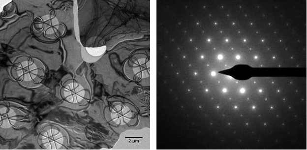
times and improved spatial resolutions can be achieved. The figures below show
(left panel) a bright-field TEM image of a single C36H74
crystal displaying dramatic and symmetric bend contours where the material has
solidified over pores in the support grid, and (right panel) a parallel-beam diffraction
pattern obtained from the same crystal wherein Bragg spots at relatively large
scattering angles are observed corresponding to ~1 Å distances in real space.
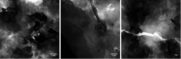
Upon addition of 10 wt% EVA to solutions of C36H74
in decane and preparation of TEM specimens via the drop-cast method, clear
differences in morphology are observed in bright-field images (see the
three-panel figure below for representative images). Most notably, the
long-range order observed in the pristine single crystals is no longer
present. Instead, the material consists of smaller, discrete regions on the
order of one micrometer with little to no crystallinity present, as verified
with parallel-beam diffraction. Further, the material forms into clumps of
varying thickness, as seen via thickness contrast in the images. These results
suggest the EVA is disrupting the chain ordering of C36H74
at the molecular level which results in the formation of amorphous globules on
the micrometer scale. Despite being in the solid state, these globules still retain
the appearance of having little to no inter-particle interactions beyond weak
forces. From this, it is expected that particle agglomeration is reduced and growth
of large paraffin crystals and/or globules in solution is hindered due to EVA
incorporation and disruption of chain ordering.
With initial preparation and characterization studies
complete, the second phase of the project will focus on time-resolved studies
of chain ordering and globule formation with UEM. In general, typical UEM
experiments will involve rapid heating of a thin n-paraffin wax crystal in
situ with a femtosecond or nanosecond laser pulse. A discrete packet of
photoelectrons generated in the gun region of the microscope with a second
laser pulse will be used to probe the specimen at some well-defined time after
excitation. Structural information is encoded on the probing electron
wavefronts in the femtosecond/nanosecond packet, and dynamics will be visualized
by varying the relative time of arrival of the pulses in a pump/probe
configuration. Atomic-scale structural dynamics will be followed with parallel-beam
electron diffraction (Bragg spot intensity, position, and width), while
processes occurring on larger scales will be mapped via bright- and dark-field imaging,
both with femtosecond to nanosecond temporal resolution. Importantly, the beam
currents used in UEM experiments are much lower than those used in standard TEM
experiments. Further, the emission process can be controlled with respect to
both time and total number of electrons per packet. In this way, we
hypothesize that beam damage can be further mitigated and, combined with
specimen cooling to 90 K, the effects of rapid laser heating can be isolated
and quantified.
The ACS PRF DNI has enabled the research direction
described above by providing student support early in the career of the PI. We
view this funding as a form of seed money for generating much-needed
preliminary results on the challenging problem of high spatial and temporal
resolution studies of soft matter and, potentially, biological structures with
UEM. We envision the acquisition of these results, enabled by the PRF DNI, as
forming an important part of future grant submissions to government funding
agencies. Further, and more importantly, the funding provided by the PRF has
enabled critical support for graduate students early in their scholarly
pursuits, free of the worry of limiting instrument time, purchase of supplies,
changing projects, etc. The impact of such funding early in the career of a PI
starting a challenging research program cannot be overemphasized. The work
that has been enabled by the PRF DNI is certain to have a lasting impact on our
research group for years to come.
 printer friendly