Reports: UNI553617-UNI5: Engineering of Interfacial Barrier Properties to Reduce Oxidation in Emulsions

Rohan V. Tikekar, Drexel University

MATERIALS AND METHODS

Synthesis of Si-ε-polylysine (EPL) hybrid microparticles: Eight percent (w/v) LUDOX®HS-30 (anionic) colloidal silica solution and 8% EPL (cationic) solution were prepared in ultrapure water. To prepare EPL coated silica hybrid microparticles, the anionic silica nanoparticle solution and cationic EPL solution were mixed in 1:1 volume proportion. The total volume of the aqueous phase was 25mL. The solution was stirred for 30 minutes to facilitate hybrid microparticles formation. The pH of aqueous phase was adjusted to 6.  4% LUDOX®HS-30 solution adjusted to pH of 6 was also prepared to synthesize Si colloidosomes.

Synthesis of colloidosomes: The lipid phase was prepared by mixing BODIPY®665/676 dye (3 ug/g lipid phase prepared in chloroform) with olive oil (5% w/v aqueous phase). Coarse colloidosomes were prepared by mixing aqueous phase containing Si-EPL hybrid microparticles and lipid phase using a high speed disperser set at 8,000rpm for 2min. The size of colloidosomes was further decreased by sonicator at 60% amplitude for 3min. 0.1% sodium azide was added to prevent microbial growth. Si colloidosomes were prepared in the exact same manner except the aqueous phase consisted of anionic 4% LUDOX®HS-30.


Characterization of colloidosomes: Particle size and ζ-potential of the silica nanoparticles, Si-EPL hybrid microparticles and colloidosomes were measured. The SEM images of Si-EPL hybrid microparticles were collected by a Zeiss Supra 50VP Field Emission Scanning Electron Microscope.


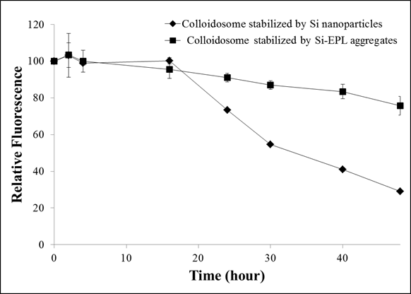
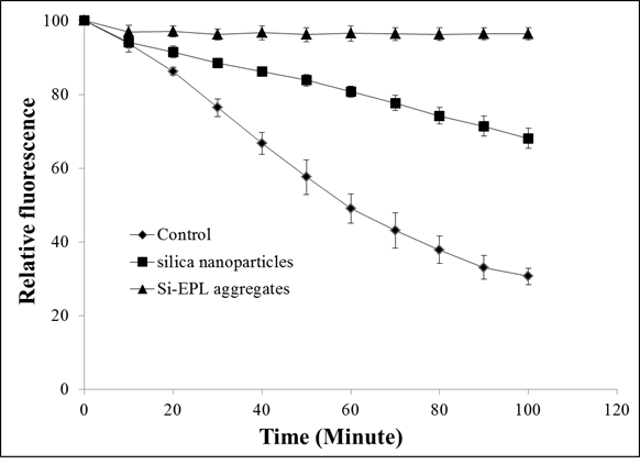
Measurement of barrier properties of Colloidosomes:  2mL of colloidosome sample was mixed with 2mL of 40mM AAPH solution and thoroughly mixed. The control samples were prepared by mixing 2mL of colloidosomes with 2mL of water. 200 uL of the samples were placed in the plate-reader to measure the fluorescence intensity of peroxyl radicals sensitive dye to peroxyl radicals generated in the aqueous phase by AAPH as a function of time. The values were obtained every few hours for a total period of 40 hours. The excitation and emission wavelengths were 620nm and 675nm respectively. The relative fluorescence intensity was calculated using equation 1:

    (1)

where, It AAPH is the fluorescence intensity of the sample after ‘t’ minutes upon exposure to AAPH, I0 AAPH is the fluorescence intensity of the sample immediately after adding AAPH, It control  is the fluorescence intensity of control after ‘t’ minutes, I0 control is the fluorescence intensity of control immediately after adding AAPH.


RESULTS AND DISCUSSION


Characterization of Si-EPL hybrid microparticles and Colloidosomes: The average particle size of silica nanoparticles and Si-EPL hybrid microparticles was 11.6±0.56 nm and 414±44.4 nm 
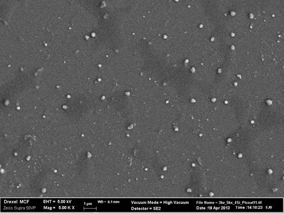
respectively. The SEM image of Si-EPL hybrid microparticles is shown in Figure 1. Large non-uniform particles formed due to aggregation of anionic silica and cationic EPL could be observed clearly. The average particle size obtained based on the image analysis of 19 hybrid microparticles was approximately 339±63 nm. Thus the average particle sizes measured by DLS and SEM were approximately similar. The average droplet sizes colloidosomes stabilized by silica nanoparticles and silica-EPL microparticles were 118±55.3 nm and 679.5±47.5 nm respectively.  These results demonstrate that formation of polymer silica hybrid complex significantly (p<0.05) increased the size of the silica particles and the resulting colloidosomes.          

  Effect of Hybrid microparticles on Barrier Properties of Colloidosomes: Figure 2 shows the loss of relative fluorescence intensity of BODIPY® 665/676 intensity encapsulated within Si and Si-EPL stabilized colloidosomes upon exposure to AAPH (20mM) in the aqueous phase. . The results show a significantly higher (p<0.05) rate of loss of fluorescence for colloidosome stabilized by silica nanoparticles as compared to colloidosome stabilized by Si-EPL hybrid microparticles. After 24 hours of incubation, the relative fluorescence in both colloidosomes began to decrease, but the rate of decrease in Si-EPL colloidosome was significantly lower. After 40 hours incubation with AAPH, the average relative fluorescence intensity in Si-EPL stabilized colloidosomes had decreased by only 17%, while the fluorescence intensity of colloidosomes stabilized anionic silica nanoparticle decreased by more than 60%. These results suggest that the rate of permeation of peroxyl radicals in the Si-EPL stabilized colloidosomes was significantly (p<0.05) lower compared to Si stabilized colloidosomes.  A mechanism that may be responsible for the observed effects is the ability of EPL to chemically interact with peroxyl radicals and prevent their permeation across the interface. In order to test this hypothesis, we evaluated the rate of fluorescence quenching of fluorescein by peroxyl radicals generated from 20 mM AAPH in distilled water in the presence or absence of anionic Si nanoparticles and Si-EPL hybrid microparticles. Fluorescein is a water soluble fluorescent dye that loses its fluorescence intensity upon exposure to hydroxyl and peroxyl radicals. It is important to note that, in this experiment, only Si-nanoparticles and Si-EPL hybrid microparticles dispersed in the aqueous phase were used without the presence of oil phase. Figure 3 shows the rate of loss of fluorescence signal intensity of the fluorescein dye both in the absence or presence of these particles. The relative fluorescence intensity decreased by approximately 80% after 100 minute of exposure in control. In presence of anionic silica nanoparticles, the rate of loss of fluorescence decreased significantly and approximately 30% decrease in fluorescence intensity was observed after 100 minute of exposure, indicating silica nanoparticles preferentially interacted with peroxyl radicals over fluorescein. However, in presence of SI-EPL hybrid microparticles, there was only a 5% decrease in the relative fluorescence after 100 minutes of exposure to peroxyl radicals. This result clearly indicates that Si-EPL aggregates were much more effective in chemically interacting and quenching peroxyl radicals compared to anionic Si- nanoparticles. This could be attributed to reducing ability of EPL polymer. Another mechanism that may also be responsible for lower peroxyl radical permeation in Si-EPL stabilized colloidosomes is higher interfacial thickness.


Figure 1: Scanning electron microscopy images of Si-EPL Hybrid microparticles.


Figure 2: Measurement of the decay rate of peroxyl radical sensitive dye encapsulated in colloidosomes stabilized by anionic silica nanoparticles and Si-EPL hybrid microparticles.


Figure 3: Decay rate of fluorescence from fluorescein in the absence and presence of silica nanoparticles and Si-EPL hybrid microparticles.