Reports: UNI1053401-UNI10: Probing Phonons in Low-Dimensional Thermoelectric Materials by Raman Spectroscopy
Rui He, PhD, University of Northern Iowa
In this research, we
used Raman spectroscopy to study laser induced oxidation, vibrational, and
optical properties of stoichiometric and non-stoichiometric Bi2Te3
nanoplates (NPs). Bi-Te nanoplates with different thicknesses were grown by low
pressure vapor transport method by our collaborators (Dr. Xuan Gao’s group) at
Case Western Reserve University. In our studies, we used a Horiba Labram Raman
microscope system and focused the laser to a spot with a diameter of ~1 μm.
Other experimental techniques-atomic force microscopy
(AFM), energy-dispersive X-ray spectroscopy (EDS), and Auger electron
spectroscopy (AES)-were also used to characterize the
sample properties.
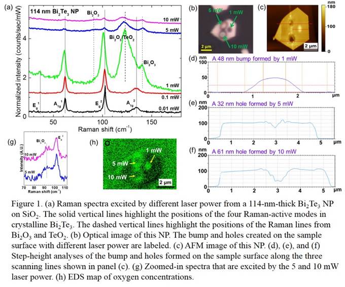
In relatively thick
(>50 nm) stoichiometric Bi2Te3 NPs, we found that the
crystalline structure is stable and sample surfaces do not show any damage under
low laser power (much lower than 1 mW) irradiation. Raman spectra show four
characteristic peaks from crystalline Bi2Te3 structures
(as highlighted by the four solid vertical lines in Fig. 1(a)). As the laser
power increases to an intermediate level (~1 mW), the NPs are oxidized and form
bumps on the surfaces (see the optical image in Fig. 1(b), and the AFM image and
profile analysis in Figs. 1(c) and (d), respectively) possibly due to expanded
crystal lattice. The oxidation is revealed by the emergence of Bi2O3/TeO2
Raman lines (highlighted by dashed vertical lines in Fig. 1(a)) and the
increase of oxygen concentration in an EDS map (Fig. 1(h)) of the NP. Further
increase of laser power not only causes oxidation (see Figs. 1(a) and (g) for
Bi2O3/TeO2 Raman lines, Fig. 1(b) for the
optical image, and Fig. 1(h) for EDS map of oxygen concentration), but also burns
holes on the sample surface. The AFM image and step-height analysis in Figs.
1(c), (e), and (f) show that the higher the laser power, the deeper the holes
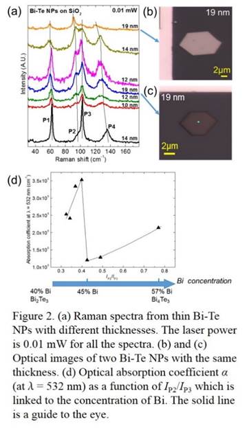
are.In NPs that are thinner
than 20 nm and grown by the same method, we found that the Raman modes are
different from those of stoichiometric Bi2Te3 crystalline
structures and are consistent with those from Bi-rich Bi-Te materials. From the
relative intensity between P2 and P3 modes (see Fig. 2(a)), we estimate that the
Bi concentration is between 40-57% in our thin NPs. We confirmed the
stoichiometries of two thin NPs by AES, which showed excellent agreement with
those estimated using Raman method. We found that the laser induced oxidation
is not prominent in these non-stoichiometric thin NPs. We also found that the
optical absorption of the thin NPs strongly depends on their stoichiometry. NPs
with the same thickness but different stoichiometries show very different color
contrast compared to the SiO2 substrate (see Figs. 2(b) and (c)). We
estimated the optical absorption coefficient of thin Bi-Te NPs with different
stoichiometries by comparing the intensities of the Si Raman lines when the
laser is directly incident on the substrate and after it penetrates the NPs. Figure
2(d) shows the optical absorption coefficient as a function of the relative intensity
between P2 and P3 and as a function of Bi concentration.Our results show that thin
Bi-Te NPs grown by the low pressure vapor transport technique show various stoichiometries
that differ from Bi2Te3, and that the optical properties
of these NPs is strongly influenced by their stoichiometry. Therefore, controlling
the stoichiometry in the Bi-Te NP growth is important for their thermoelectric,
electronic, and optical device applications. Details of these findings are to
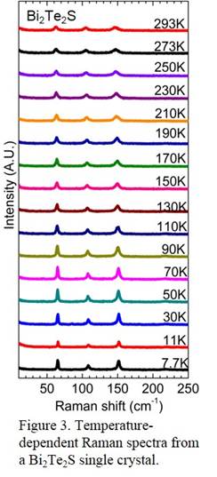
be published in Nano Research. A new closed cycle
optical microscopy cryostat was installed in the PI’s lab in June 2014. This
equipment, together with the Raman microscope system, has enabled variable
temperature optical microscopy studies of diverse materials. Figure 3 shows
preliminary data of temperature dependent Raman spectra from a Bi2Te2S
single crystal (samples were provided by Dr. Yong Chen’s group at Purdue
University). Students will analyze the data under the PI’s supervision in the
coming academic year.This PRF grant has
allowed the PI to explore a new research area that she has initiated since she
started her career as an assistant professor at the University of Northern Iowa
(UNI). The research has stimulated vigorous internal (within the PI’s
department) and external collaborations that connect UNI, a predominantly
undergraduate institution, with Case Western Reserve University and Purdue
University, Ph.D.-granting research institutions. Five papers were published
during this PRF grant support period (see Publication list) and one paper was
recently accepted for publication in Nano Research.The research projects
enabled by this grant open up new opportunities for our undergraduate students
to improve their preparation for graduate school and the STEM workforce. Four
physics major undergraduates (Casie Means-Shively, Courtney Keiser, Chao Ji,
and Zhipeng Ye) were supported by the PRF grant and participated in the research
activities. A new student, Heidi Anderson, started her research in August 2014
and is also supported by the PRF grant. Courtney Keiser and Zhipeng Ye’s
research in the PI’s group was supported by PRF since fall 2013. They are
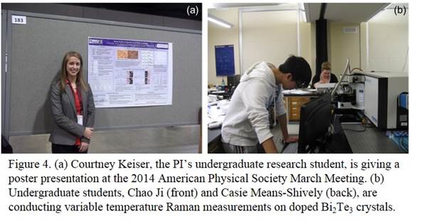
coauthors on two papers published during this grant active year. Courtney
Keiser attended the 2014 American Physical Society March Meeting held in Denver
Colorado and presented the research on Bi2Te3 nanoplates
at the meeting (see Fig. 4(a)). Chao Ji and Casie Means-Shively joined the PI’s
group in summer 2014 and participated in the research of temperature dependent
Raman studies of phonon properties of doped Bi2Te3
crystals (see Fig. 4(b)). This research experience offered by the PRF grant has
stimulated our students to pursue advanced education in graduate school to
further their education.
 printer friendly