57th Annual Report on Research 2012 Under Sponsorship of the ACS Petroleum Research Fund
Reports: DNI350270-DNI3: Molecular Mechanism of Methane to Methanol Conversion in Soluble Methane Monooxygenase
Serena DeBeer, PhD, Cornell University
Methane monoxygenases (MMOs), found in methantrophic bacteria, are Nature's catalysts for the controlled oxidation and functionalization of strong C-H bonds. The soluble form of MMO utilizes a binuclear iron active site that reacts with dioxygen proceeding through a mu-peroxo-Fe(III)Fe(III) species (MMO-P) and then an Fe(IV)Fe(IV) species (MMO-Q). The later is responsible for methane hydroxylation, however, its structure remains a subject of debate. The present research is focused developing X-ray spectroscopic methods, which will allow for a novel means to characterize reactive intermediates. During the last year, we have focused on the x-ray emission (XES) characterization of binuclear iron models, with close correlation to theory. We have also initiated new high-resolution fluorescence detection x-ray absorption spectroscopic (HERFD-XAS) studies, which should provide further insight into the electronic structures of iron intermediates.
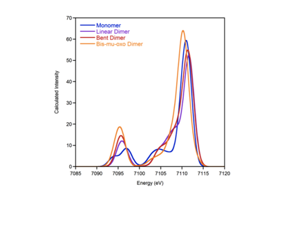
Studies of binuclear model complexes by XES. During the current grant period, we have focused on understanding the valence to core XES (V2C XES) of binuclear iron complexes, with varied oxygen bridging motifs. The V2C XES results from an electron in a ligand-based molecular orbital refilling an iron 1s core hole, created by photoionization. As such, the method is very sensitive to ligand identity and ionization potential. In order to test the sensitivity of this method to the bridging motif in binuclear iron complexes, we have studied a series of twelve iron dimers, which have included mono-mu-oxo, bis-mu-oxo and hydroxo species. A representative sampling of these data are shown in Figure 1. Interestingly, based on the current literature understanding, one would expect the intensity of the oxygen 2s to iron 1s satellite feature at ~7095 eV to increase in intensity with decreasing iron-oxygen bond length. As the figure shows, however, there is no clear correlation based on metal ligand distance. Rather, we find that there is a pronounced contribution from Fe-O-Fe angle, with the greatest intensity resulting when the Fe-O-Fe angle approaches 90 degrees. Conversely, the satellite peak is entirely absent, when the angle approaches 180 degrees. Thus the spectral intensity mechanism can be understood in terms of a simple Walsh diagram for mixing of the oxygen 2s with the iron np orbitals. This effect can also be reproduced computationally (Figure 2). These results demonstrate a dependence of the spectra on the core geometry, which was not previously known. These data also provide markers for studies of the enzymatic intermediates. We are currently preparing a manuscript, which describes our studies on the binuclear iron models. In addition, we continue to pursue XES measurements on the enzymatic intermediates, though low concentrations and issues with photoreduction have proven problematic.
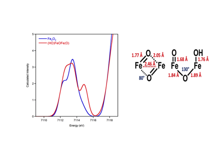
Studies of high-valent intermediates by HERFD XAS. In conjunction with our XES studies, we are also pursuing HERFD XAS studies. HERFD XAS utilizes a high resolution crystal spectrometer to detect the K-alpha fluorescence, resulting in greatly improved spectral resolution, thus enhancing the information content of the data. We have already shown that TDDFT approaches can predict standard Fe K-edge XAS spectra with good reliability. We are currently extending these methods to HERFD XAS data. In addition, we have calculated spectra for the enzymatic intermediates. Our preliminary calculations show that these data should allow us to distinguish the two proposed core structures for MMO-Q – i.e. the bis-μ-oxo diamond core (FeIV2O2) or an open core (HO-FeIV-O-FeIV=O) (Figure 3). The HERFD XAS measurements on MMO-Q are planned at the European Synchrotron Research Facility in January 2013.
The work funded by this PRF grant has been essential in establishing my research groups understanding of XES spectra and their potential applications to enzymatic intermediates. This work forms a substantial portion of one post-doctoral fellows research, as well as one Ph.D student.
Figure 1. Representative binuclear model complexes studied by V2C XES.
Figure 2. Calculated V2C XES as a function of Fe-O-Fe angle.
Figure 3. Calculated HERFD XAS for the two proposed core structures of MMO-Q.


