Reports: DNI651233-DNI6: Fundamental Antenna-Receiver Interactions in Metal-Based Light-Harvesting Nanostructures Studied by Magneto-Optical Spectroscopy
 printer friendly
During our first ACS funding year, we have made significant research progress. Upon completion, my research group will possess the experimental and theoretical expertise to probe state-specific interactions between nanoparticle plasmon resonances and molecular electric-dipoles. The predictive understanding of plasmon-electric-dipole interactions arising from this fundamental research program may result in enhanced solar photo-conversion efficiency. In particular, our use of plasmon-enhanced magneto-optical spectroscopy can impact several areas of chemistry by providing insight into nanostructure oxidation and spin state. Progress toward these goals in three key areas are highlighted: i) single-particle nonlinear optical (NLO) spectroscopy for probing nanoscale surface electromagnetic fields, ii) implementation of continuous polarization variation (CPV) analysis to quantify nanoscale surface electric fields and magnetization components, and iii) the assembly and characterization of molecular-bridged nanoparticle dimers.
I. Single-particle Polarization-resolved NLO Spectroscopy
![Text Box:
Figure 1. Inter-particle-gap-to-diameter-dependent (D/2r) SHG depolarization ratios. Experimental SHG depolarization ratios, [I2w(x)] /[I2w(y)], (black square) as a function of D/2r ratio. Red dots represent the numerically simulated depolarization ratio (Ex/Ey) at 800 nm.](images/abimages/Paper_11966_abstract_17876_0.png) The
first research stage involved development and characterization of a microscope
capable of second harmonic generation (SHG) measurements at the single-particle
level. Single-particle measurements are necessary because metal nanoparticles
are inherently heterogeneous. SHG, a second-order NLO technique, was chosen for
nanoscale surface field investigation because the intensity of these
measurements increases as the 4th power of local field strength.1
SHG measurements involve focusing the fundamental output of a mode-locked laser
to the sample plane using an aspheric (NA = 0.5) lens. Interactions between the
fundamental wave and the sample result in SHG, which is isolated from laser light
using a series of filters. Signal is detected by a photomultiplier tube
operated in photon-counting mode. Our home-built microscope can also collect
bright-field imaging data, allowing for spatial correlation of SHG data with
specific nanoparticle structures that have been deposited onto microscope
slides that are alpha-numerically indexed. Regions of experimental significance
are determined from the bright-field image and then subjected to structural
analysis via electron microscopy.
The
first research stage involved development and characterization of a microscope
capable of second harmonic generation (SHG) measurements at the single-particle
level. Single-particle measurements are necessary because metal nanoparticles
are inherently heterogeneous. SHG, a second-order NLO technique, was chosen for
nanoscale surface field investigation because the intensity of these
measurements increases as the 4th power of local field strength.1
SHG measurements involve focusing the fundamental output of a mode-locked laser
to the sample plane using an aspheric (NA = 0.5) lens. Interactions between the
fundamental wave and the sample result in SHG, which is isolated from laser light
using a series of filters. Signal is detected by a photomultiplier tube
operated in photon-counting mode. Our home-built microscope can also collect
bright-field imaging data, allowing for spatial correlation of SHG data with
specific nanoparticle structures that have been deposited onto microscope
slides that are alpha-numerically indexed. Regions of experimental significance
are determined from the bright-field image and then subjected to structural
analysis via electron microscopy.
The extent of SHG sensitivity to local surface fields was examined by analyzing the depolarization ratios determined for a series of SGN dimers with different interparticle-gap-to-particle-diameter (D/2r) values. SGN dimers were chosen as model system because their particle diameter- and distance-dependent optical properties are understood.2 Depolarization ratios decayed as an exponential function of D/2r (Fig. 1), consistent with plasmon-ruler expectations. Numerical simulations of the SGN dimer electric field reproduced the depolarization values. To describe these data fully, it was necessary to account for both absorption and scattering contributions to the SGN dimer optical properties, which we detailed in a full article. The strong agreement between calculated and experimental SHG depolarization ratios demonstrates the capacity of single-particle SHG measurements to quantify nanoparticle surface fields.
II. Continuous-polarization-variation-detected SHG.
A significant advantage of SHG measurements over emission and linear scattering techniques is that the signal is described by the sample's nonlinear susceptibility. A general expression describing the experimentally measured SHG intensity I(2w) as a function of the polarization of the fundamental wave follows:
![]()
 S(f) and P(f)
are the fundamental polarization states in the s and p laboratory frames; F, G,
and H are nonlinear susceptibility components. F corresponds to
electric-dipolar contributions, G to magnetic-dipolar contributions, and H to a
linear combination of the two. Therefore, SHG measurements conducted using
continuous polarization variation inform on nanoparticle surface field
symmetry.
S(f) and P(f)
are the fundamental polarization states in the s and p laboratory frames; F, G,
and H are nonlinear susceptibility components. F corresponds to
electric-dipolar contributions, G to magnetic-dipolar contributions, and H to a
linear combination of the two. Therefore, SHG measurements conducted using
continuous polarization variation inform on nanoparticle surface field
symmetry.
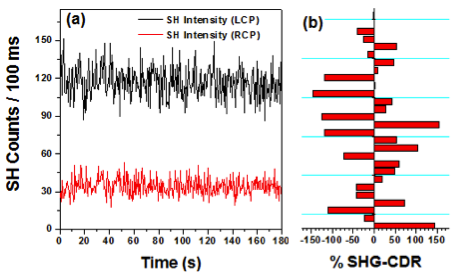
To demonstrate our ability to collect CPV-resolved data, SHG-detected circular dichroism measurements were made selecting the incident polarization state as either right or left circularly polarized and quantifying the differential SHG response (Figure 2a). The data demonstrate a large, unambiguous difference in the SHG signal upon switching from right to left circularly polarized incident light, indicating a chiral plasmon field. The extent of this chirality was determined using the SHG circular-difference ratio (SHG-CDR):
![]()
The SHG-CDR can assume values between 0 and 2. In a study of several SGN dimers, we observed non-zero CD values for many (but not all) structures, with a large range of experimentally determined SHG-CDR values arising from different SGN dimers. Responses from twenty-seven different structures (summarized in Figure 2b) demonstrate that a high level of structure specificity can be achieved using single-particle NLO measurements.
An important result from this research stage is the use of colloidal nanoparticles to amplify circularly polarized electromagnetic fields, which represents a major step toward achieving spatially resolved single-particle magneto-optical measurements. This research will enhance the understanding of electronic structure of molecules near nanoparticle surfaces and provide descriptions of interactions between plasmon modes and specific molecular states. In addition to achieving the proposed research goals, synthesis and assembly of nanoparticle dimers and development of advanced experimental facilities suited for plasmon-enhanced magneto-optical spectroscopy will allow the PI to achieve his long-term research and education objectives.
III. Molecularly-bridged SGN dimers
Progress has also been made in the preparation and characterization of iron porphyrin-bridged SGN dimers. SGN dimers are formed by electrostatic interactions between the porphyrin and negatively charged gold nanoparticles; the N-CH3+ groups of the iron porphyrin located in the interparticle gap between dimers interacts with citrate molecules on the SGN surface. Surface-enhanced Raman data, which exhibit prominent peaks at 660, 775, 865 and 1180 cm-1 support this conclusion. These signals arise from vibrations of N-CH3+ groups positioned near the nanoparticle surface when the porphyrin binds the metal in an edge-on geometry.3 During the next year of ACS support, we will incorporate our NLO microscope into a superconducting magnet, allowing for plasmon-enhanced magneto-optical imaging. Iron porphyrins, whose MCD spectra have been described in detail, will be used as the model system.
In addition to providing the PI with experimental infrastructure to perform fundamental plasmon-enhanced magneto-opitcal measurements on light-harvesting nanostructures, the research effort has provided training for three students (2 graduate, 1 undergraduate): Anne-Marie Dowgiallo prepared and characterized nanoparticle assemblies; Jeremy Jarrett developed SHG facilities; Patrick Herbert also made significant contributions.
1. R.F. Boyd, Nonlinear Optics, Academic Press, San Diego, CA, USA, 1992.
2. B.M. Reinhard et al. Nano Lett. 2005, 5, 2246.
3. S. Rywkin et al. Langmuir, 2002, 18, 5869.