AmericanChemicalSociety.com
Reports: UFS 49374-UFS: Abundance and Stable Carbon Analysis of Phospholipid Fatty Acids in Association with Methane Hydrates in the Arctic Ocean
Thanks to the PRF UFS grant program, I had the opportunity to participate in the Methane In The Arctic Shelf (MITAS) research cruise in the Arctic Ocean. The expedition explored the geophysical, biological and geochemical properties of methane hydrate formations in the Beaufort Seas. I collected sediment samples from cores retrieved off shore of Prudoe Bay, Alaska. My goal is to quantify the phospholipid fatty acids (PLFAs) and their carbon isotope values found within these samples. This information should help elucidate the biogeochemical processes of Arctic gas hydrate formation and destruction.
Gas hydrate deposits along continental margins and in Arctic regions contain a large quantity of methane.1 The Arctic Ocean represents 1% of total ocean volume, however, its depth of 1361 meters allows continental slopes and rises containing thick sediment successions containing considerable organic-rich natural gas source beds and permeable sandy beds that concentrate gas hydrates.2 Added to these coastal hydrates, are permafrost hydrates developed in geological traps during the glacial episode. This suggests the Arctic Ocean is a key region for new energy exploration.3
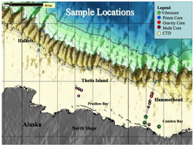
The MITAS research cruise focused on the section of the Arctic Ocean known as the Beaufort Sea (Figure 1) which extends to the northeast from Point Barrow, Alaska to the northeast of Prince Patrick Island, northward toward Banks Island and westward to the Chukchi Sea. The average depth of the Beaufort Sea is 1,004 m. The sediment character in this region is controlled by dispersal and re-suspension of river-borne sediments ice scouring, and coastal erosion and retreat.4 In the eastern Beaufort Sea, from the Mackenzie River up to approximately130-106 tones/yr. To the west of the Mackenzie River system, along the western Beaufort Shelf, sediment delivery results from numerous arctic river systems including the Colville River.5
Figure 1. Sample Locations Phospholipid fatty acids are the central component of bacterial cell
membranes. When organisms die,
PLFA begins to quickly degrade, so sediment PLFA concentrations should
correlate with only the total viable bacterial populations.6,7
Specific PLFA structures and ratios vary by type or bacterial group.8,9
Finally, the stable carbon isotope ratio of PLFAs can provide insight into
microbial carbon sources, separating archae involved in the anaerobic methane
oxidation and bacteria responsible for sulfate reduction. I am in the process of using lipid biomarkers and stable carbon isotope
measurements to assess the microorganisms that drive carbon cycling in Artic
Ocean gas hydrate systems. Subsamples of sediment cores taken during the Arctic
expedition have been frozen at -80 °C until analysis. PLFAs are being Soxhlet extracted using a
modified Bligh Dyer method.10 A series of varying polarity solvents
will be used on solid phase columns to isolate PLFAs from the bulk extract.11
The isolated PLFAs will be converted to Fatty acid methyl esters (FAMEs) via a
mild alkaline methanolsis.12,13 An aliquot of the FAME will
subsequently be converted to their dimethyl disulfide adducts to determine the
bond positions of the monounsaturated FAMEs.12,14 The FAMEs will be quantified using an Agilent 6890 GC equipped with a 60
m HP-88 capillary column specific for FAME separation and a 5973 mass
spectrometer. Compound specific carbon isotope values of the FAMEs will be
determined on a Finnigan DELTA Plus XP IRMS interfaced with a GC/C-III to a
TRACE GC ULTRA with a 60 m HP-88 column. Measured values will be corrected for
the addition of carbon during derivatization.15 Compound
identification will be based on relative retention times against known
standards and retention times. Quantification will be based on the addition of
the methyl esters of 13:0 and 20:0 PLFA used as internal standards. This sabbatical provided me with new opportunities to investigate energy
sources, conduct oceanographic research as well as generating new information
on the formation of Arctic gas hydrates.
Undergraduate in my lab are testing the use of DRIFTS-FTIR and XRF to
glean additional information from the samples. Additionally, several new projects are being planned or have
been initiated as a result of this research. Collaborations with oceanographers at North Carolina State
University and Texas A&M have begun to analyze my samples for Humic
Substances and lignin content, respectively. I've also been invited to participate in a hydrate
exploration cruise in the Gulf of Mexico as well as in the Kara Sea during
2011. References 1. Kvenvolden,
K. A. 1999. Proc.Natl.Acad.Sci.U.S.A. 96: 3420-3426. 2. Max, M.D.
& Lowrie, A. 1993. Natural gas hydrates: Arctic and Nordic Sea potential.
In: Vorren, T.O., Bergsager, E., Dahl-Stamnes, ¯. A., Holter, E., Johansen, B.,
Lie, E. & Lund, T.B. Arctic Geology and Petroleum Potential, Proceedings of
the Norwegian Petroleum Society Conference, 15-17 August 1990, Tromsz, Norway.
Norwegian Petroleum Society (NPF), Special Publication 2 Elsevier, Amsterdam,
27-53. 3. Collett, T.
S., 2003. Natural Gas Hydrate as a Potential Energy Resource. In: Max, M. D..
(ed.), Natural Gas Hydrate in Oceanic and Permafrost Environments. Kluwer
Academic Publishers, Dordrecht, 123-136. 4. Carmack, E. C. and R. W.
MacDonald. 2002. Arctic. 56:29-45. 5. Dunton, K.H., Weingartner,
T., and Carmack, E.C., 2006. Progress in Oceanography 71:362–378. 6. Harvey,
H.R., Rallon, R.D., Patton, J.S., 1986. Geochimica et Cosmochimica Acta 50,
795-804. 7. Mills, T.
M., Dias, R.F., Graham, D., Mandernack, K.W., 2006. Marine Chemistry 98, 197-209.
8. Green, C.T.,
Scow, K.M., 2000. Hydrogeology Journal 8, 126-141. 9. Pinkart,
H.C., Ringelerg, D.B., Piceno, Y.M., MacNaughton, S.J., White, D.C., 2002.
Biochemical approaches to biomass measurements and community structure. In:
Hurst, CJ., (Ed.), Manual of Environmental Microbiology. ASM Press,
Washington D.C. 10. Pohlman,
J.W. 2006. Doctoral Dissertation, School of Marine Science, College of William
and Mary. 241 pages. 11. Kaluzny,
M.A., Duncan, L.A., Merritt, M.V., Epps, D.E., 1985. Lipid Research 26,
135-140. 12. White,
D.C., Ringelberg, D.B., 1998. Signature lipid biomarker analysis. In: Burlage,
R.S., Atlas, R., Stahl, D, Geesey, G, Sayler, G. (Eds.), Techniques in
Microbial Ecology, Oxford University Press, New York. 13. White,
D.C., Davis, W.M., Nickels, J.S., King, J.D., Bobbie, R.J., 1979. Oecologia 40,
51-62. 14. Dunkelblum,
E., Tan, S.H., Silk, P.J., 1985.. Journal of Chemical Ecology 11, 265-277. 15. Go–i, M.A.,
and T.I. Eglinton. 1996. Organic Geochemistry 24:601-615.