AmericanChemicalSociety.com
Reports: ND7 49090-ND7: Unimolecular Polymeric Janus Nanoparticles: Synthesis, Self-Assembly and Applications
1. Project Progress
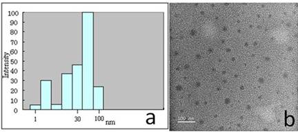
With gradual addition of water into the DMF solution of PS-b-QP2VP-b-PE, typical opalescence appeared, indicating the formation of aggregates. After dialysis to completely remove DMF, the size of micellar aggregates was determined by dynamic light scattering (DLS), which showed an average hydrodynamic diameter of 41 nm (Chart 1a). The structure of aggregates was further visualized by transmission electronic microscopy (TEM) as shown in Chart 1b. Because of the hydrophobic driving force in the aqueous solution, hydrophobic PS formed the core with hydrophilic quaternized QP2VP and PEO as corona. The concentration of the micelle solution was adjusted to 1.0 mg/mL for further complexation with ssDNA.
Chart 1. (a) Hydrodynamic diameter distributions of quaternized
PS-b-P2VP-b-PEO micelles aqueous solutions; (b) TEM image of typical micelles
from PS-b-QP2VP-b-PEO. With addition of 1.0 mL ssDNA solution (0.1 mg/mL) into 0.5 mL
triblock copolymer micelle solution (1.0 mg/mL) by syringe pump over 24 h, the
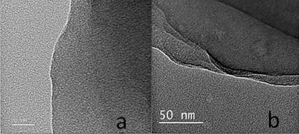
suspension started to precipitate out. The TEM image in Chart 2a clearly showed the formation of ultrathin films. Moreover,
at high amplification (Chart 2b), no
obvious phase separation was observed.
Chart 2. TEM images of
typical unltrathin films obtained after adding the ssDNA solution into the micelle solution. To understand the mechanism for the
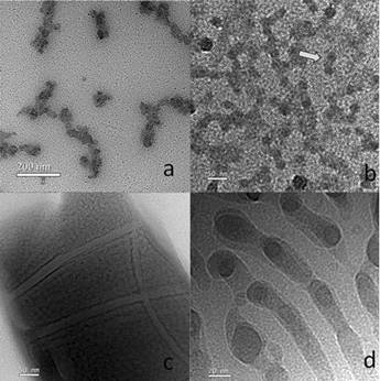
formation of ultrathin films, the intermediate sample at two hours was
investigated. Clearly, cylindrical aggregates comprised of several fused
spherical micelles are seen in Chart Thus, we speculate that after addition of ssDNA
into the PS-b-QP2PVP-b-PEO solution, one ssDNA with negative charges could permeate through the PEO layer to
complex with QP2VP with positive charges. However, the contour length of
ssDNA (~8.6 nm) is much longer than
that of QP2VP (~ 1.5 nm) and similar to the length of PEO block (~9 nm). Then, this ssDNA chain
may have two possibilities. One is to further complex with another QP2VP chain
in the same micelle and condense onto it. The other is to extend its chain outward
to further complex with another micelle. Once the second possibility happens,
two micelles link together via ssDNA,
merge and reorganize into cylindrical micelles (Chart 3b). With further aggregation, these cylindrical micelles
will merge and reorganize into the ultrathin films (Chart 3c and d).
Chart 3. TEM images for the
samples obtained at 2 h (a) without and (b) with staining using RuO4;
(c) an ultrathin film composed of the triblock copolymer and ssDNA at 24 h. (d) is a high
magnification image. As the volume ratio of ssDNA solution to the triblock copolymer
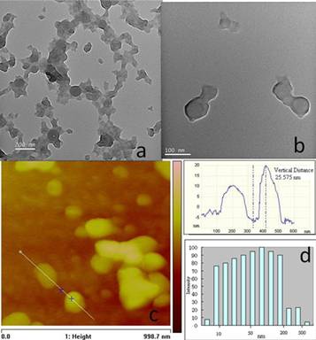
reached 3:1, the TEM observation shows that the ultrathin film disappeared gradually
and vesicular structure appeared as shown in Chart
Chart 4. The aggregates
formed at different ratios of ssDNA:PS-b-QP2VP-b-PEO after adding the DNA solution into the triblock copolymer
solution over 24 h. (a and b) TEM images and (c) AFM image; (d) Hydrodynamic
diameter distributions in PS-b-QP2VP-b-PEO/ssDNA vesicle suspension. Based on the results above, we
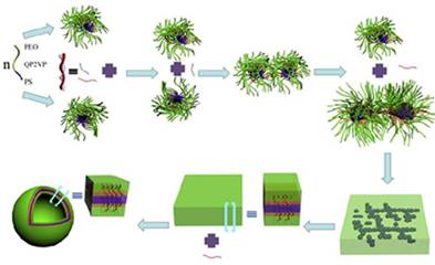
supposed the whole self-assembly process in Chart 5. In the initial stage, upon addition of ssDNA into the triblock copolymer
solution, micelles begin to link together by ssDNA to form cylindrical aggregates, then further aggregate to
form a bilayer thin film and finally form vesicular structure as the result of
the increase in volume fraction of the hydrophilic parts of the complex.
Chart
5.
The illustration of the formation of
ultrathin films and vesicles from the complexation of PS-b-QP2VP-b-PEO micelles with
ssDNA. 2. Program Impact This project has initiated a new direction of the
PI's research in the fields of supramolecular self-assembly of polymeric
particles and gene transfection, which may be viable for future grant
applications to NSF and NIH. The postdoctoral fellow has been trained to
broaden his area from organic synthesis to bio-related science and engineering,
and this is expected to be beneficial to his future professional career. High School
Student Research: During the last summer, a high school
student, Ms. Bethany Qiang from Western Reserve Acedamy, joint my group to
carry out two-week research. In this
experience, she worked with Dr. Bing Guan on TEM morphology study of the PS-b-QP2VP-b-PEO/ssDNA complexes.