www.acsprf.org
Reports: DNI548780-DNI5: Near-Field Vibrational Spectroscopy and Imaging of Chemical Species on Individual Nanoparticles During Catalytic (De)Hydrogenation
The goals of this PRF DNI project are to (1) develop a hybrid scanning probe microscopy system which combines atomic force imaging with near-field vibrational spectroscopy for local identification of surface chemistry, (2) realize catalytically-active nanoparticles with controlled shapes/compositions using solution and plasma-based methods, and (3) investigate catalytic hydrogenation reactions using the aforementioned equipment and materials. During the second year, grant funds were used to support one PhD student and purchase equipment for microscope development and catalysis experiments. The project has seen considerable advances in year two: (1) the reflection-based near-field optical microscope is operational, (2) tip-assisted Raman spectroscopy at ~10nm spatial resolution was demonstrated, (3) initial experiments to characterize single catalytic nanoparticles has been successful, (4) selective hydrogenation of acetylene with shaped Pt-Ag nanoparticles has been accomplished, and (5) microplasmas were used to synthesize metal alloy and supported catalysts for selective hydrogenation and electrocatalysis.
2. Experimental results
(a) Chemical imaging of surfaces
Our work on the development of a combined optical/atomic force
microscope for chemical imaging of surfaces using Raman spectroscopy
continued during year two. This built-from-scratch, reflection-based
instrument (Fig. 1) combines confocal optical microscopy with AFM
interrogation of the sample surface. Coupling of laser light to plasmon
modes of the tip results in enhanced optical fields in the
tip-surface gap region at distances far below the diffraction limit.
The strong optical fields tied to the tip are
used to enhance Raman scattering from molecules/nanostructures beneath
the tip; as such, the local
chemistry of the surface can be probed and imaged at high spatial
resolution using vibrational signatures. Key measurements related to
the distance scaling of Raman enhancement and Rayleigh scattering from
thin films (Si, Ge, InSb) and nanostructures (SiGe nanowires;
Pd/PdO nanoparticles) have been completed (two manuscripts will be
submitted by Oct. 30, 2011). Figure 2 shows
an AFM topography image of a 20 nm diameter SiGe nanowire on TaC, with
the accompaying line profile measured using the Raman bands of Si Ge
phonons; this data clearly shows that spectroscopic optical
interrogation and imaging at length scale approaching 10nm is feasible.
As seen, the Raman signal from the nanowire closely
follows the topography while the Rayleigh scattering remains flat
as the tip scans across the wire; this observation reflects the
plasmonic enhancement of the Raman, which can only occur when the tip
is in the optical near-field of the wire.
Raman imaging experiments on catalytic materials were also undertaken. Figure 3 shows near-field Raman at different points along an AFM scanline over a 25 nm diameter PdO/Au nanoparticle that has been coked. As seen, phonon lines from PdO (along with other carbon vibrational peaks) are only visible when the plasmonic tip is positioned over the nanoparticle. These measurements show that spatial identification/imaging of single nanoparticles and adsorbates on surfaces will be possible using near-field enhancement of Raman scattering.
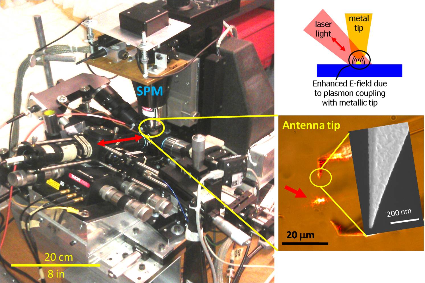
Figure 1: Reflection-mode, tip-enhanced near-field optical microscope. SPM is the scanning probe microscope; red arrows denote the confocal optical path.
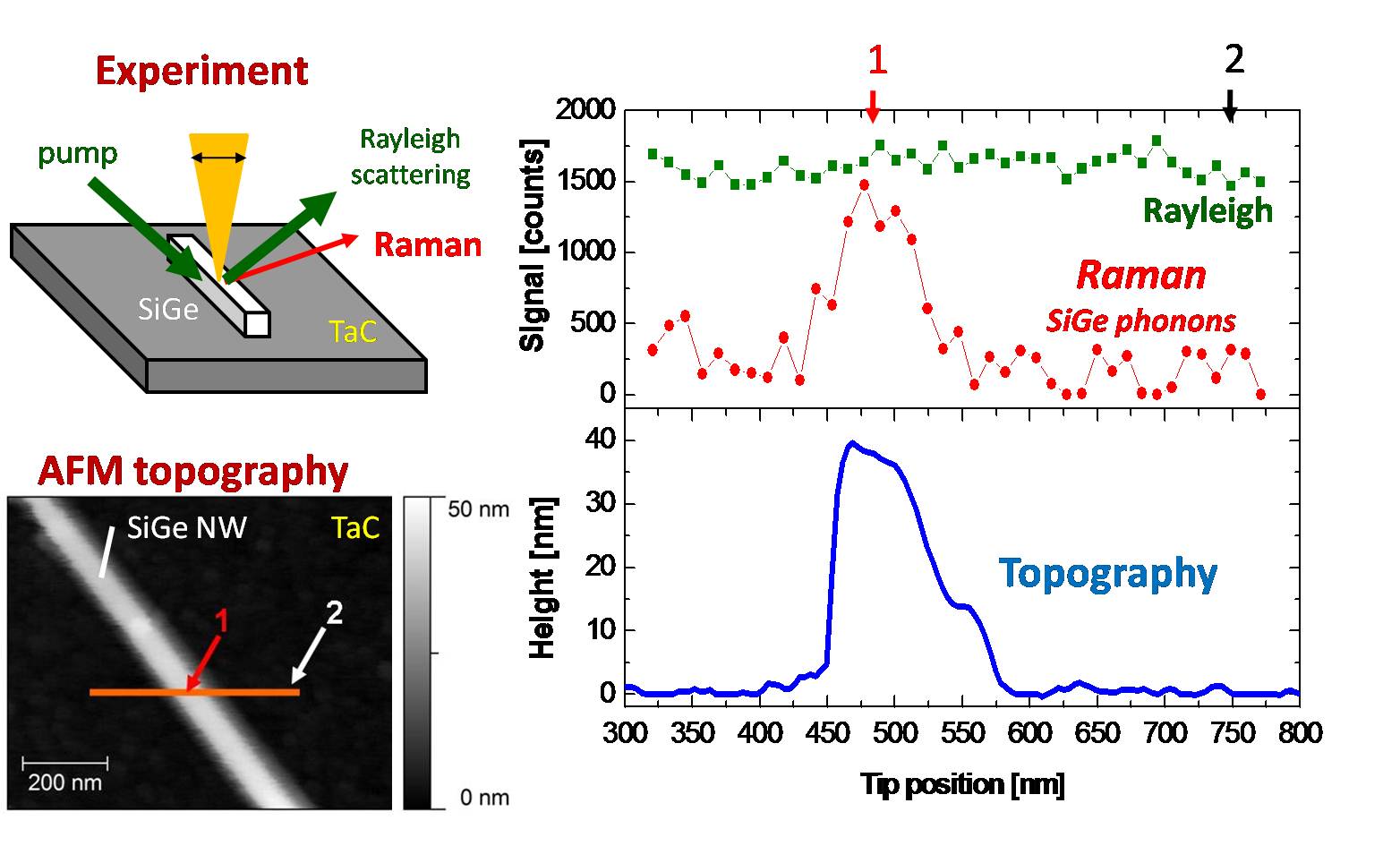 | 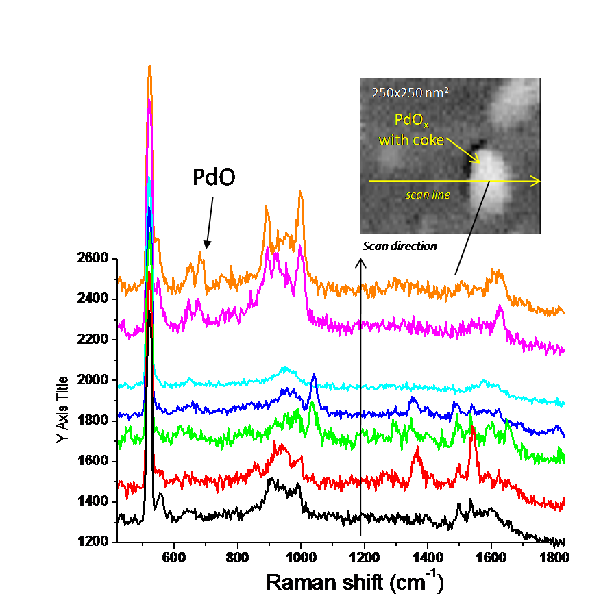 |
| Figure 2: (left) Near-field Raman interrogation of SiGe nanowire on TaC substrate with AFM topography. (right) Topography, Raman of SiGe phonons, and Rayleigh scattering for the scanline shown. | Figure 3: Raman spectra at various points along a scanline containing a 25 nm diameter PdO/Au nanoparticle. Phonon peaks from PdO appear only when the tip is over the nanoparticle (purple/orange spectra). |
(b)
Nanoparticle
synthesis & catalytic
evaluations
Our synthesis efforts to realize shaped and alloy nanoparticles via
solution and plasma-phase routes continued in year two (four
manuscripts will be submitted
by the end of 2011). Pt and PtAg nanoparticles (7-10nm cubes,
cuboctahedra, octahedra),
supported on colloidal SiO2, were tested for selective hydrogrnation of
acetylene in continuous and batch reactors.
Conclusions from this work were (1) Ag
segregates to the nanoparticle surface, (2)
nanoparticle catalysts were more active and highly selective for
C2H4 production compared to Pt black, (3) adding small amounts of Ag to
the surface
increase selectivity by >3X compared to Pt alone, (4) etching
away surface Ag lowers C2H4 selectivity (~10-20%), but
the C2H2 hydrogenation rate increases by > 500%, and (5) a
"reverse" Pt on Ag catalyst, which efficiently uses Pt, is also active
for selective hydrogenation.
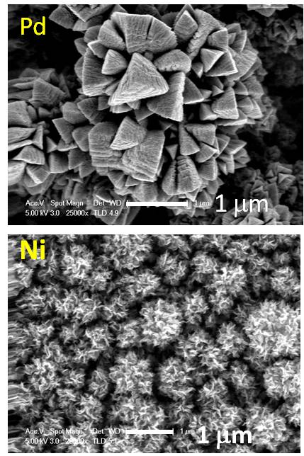
The microplasma synthesis work was extended during the last year
to realize Pd, Cu, NbOx, and PdNi / FeNi alloy nanoparticles; Pd/Ni
films; and Pd-SiO2 / Ni-SiO2 supported nanoparticles
for catalytic applications. A 100-1000X increases in particle
deposition rate from the microplasma was achieved using low
pressure operation to favor hollow-cathode operation; the
plasma jet afterglow has been used to anneal the deposited
films (i.e., the momentum of plasma ions/clusters bombarding the
substrate at high velocities - 100-300 m/s -
assists diffusion). Figure 4 shows SEM
images of nanoparticle films annealed with the jet
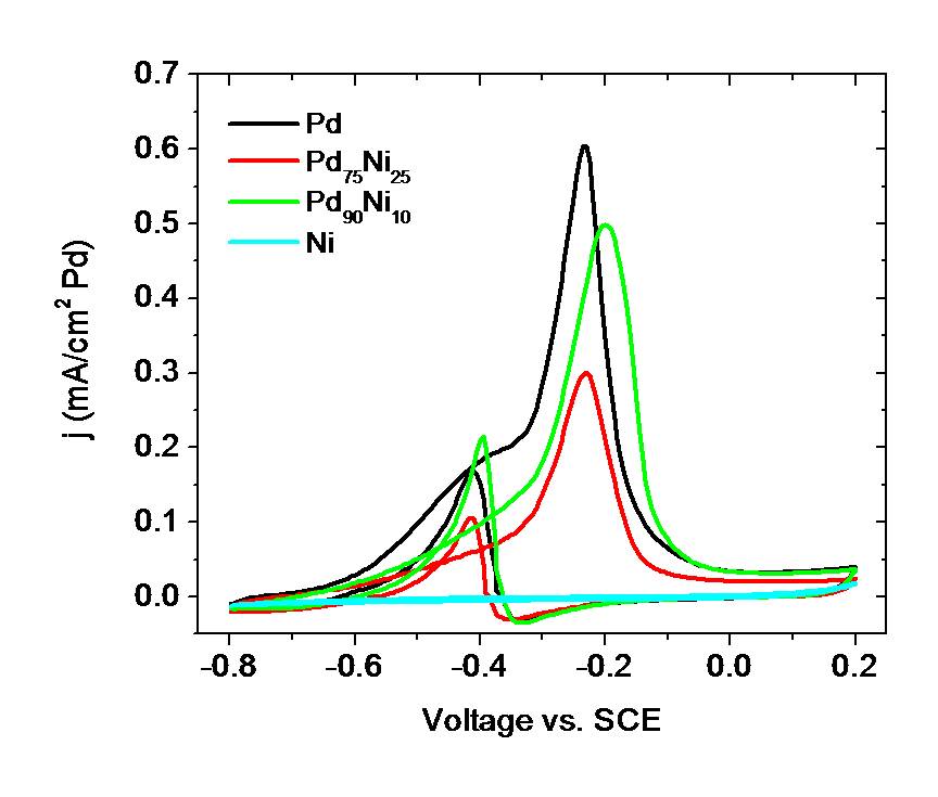
afterglow. Pd and PdNi alloy nanoparticles were tested
electrochemcially and found to be very active and stable for
methanol/ethanol oxidation in basic media (Fig. 5).
 |
  |
| Figure
4:
Pd and Ni nanostructures formed by afterglow annealing of the
nanoparticle substrate. | Figure
5:
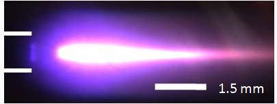
(top) Plasma jet used to form PdxNiy nanoparticles (capillary tube
noted by white lines). (bottom) Methanol electrooxidation using
different PdxNiy nanoparticle compositions. |
3.
Impact
of
research
Given the project's focus on theoretical and
experimental aspects from nanoscience, optics, and chemical synthesis,
the graduate students funded by the project are becoming
experts in the fields of scanning probe microscopy, materials
characterization, and catalysis.
These students are working in a unique laboratory setting that
provides interdisciplinary training, mentoring, and
interactions with other students/post-docs.
The PRF-DNI grant has also had a substantial impact on the PI’s ability to acquire additional funding. Preliminary results made possible by this grant were incorporated into a successful research grant from the Packard Foundation.
The equipment development/experimental work during the last year has
shown that nanoscale
chemical
imaging of catalytically-relevant surfaces and systems is possible;
in particular, the hybrid optical/scanning probe microscopy system,
synthesis work, and catalytic testing supported by this PRF grant will
allow us to probe and better understand how local effects
influence chemical reactions and molecular transformations on surfaces.