www.acsprf.org
Reports: DNI450272-DNI4: Self-Assembly of Directional Porphyrin Arrays in Water via Cyclodextrin-Based Host-Guest Interactions
Janarthanan Jayawickramarajah, PhD , Tulane University
Narrative Report Text
The overall goal of this research project is to develop well-defined and directional porphyrin arrays in water via b-cyclodextrin (b-CD)-based host-guest interactions. In particular, we had proposed to (1) construct self-assembled trimers and elucidate thermodynamic parameters of binding, (2) build linear multi-porphyrin arrays wherein array length and inter-porphyrin distances are modulated, and (3) probe energy transfer properties of the arrays. We have made significant progress on subaims 1 and 2 as outlined below.
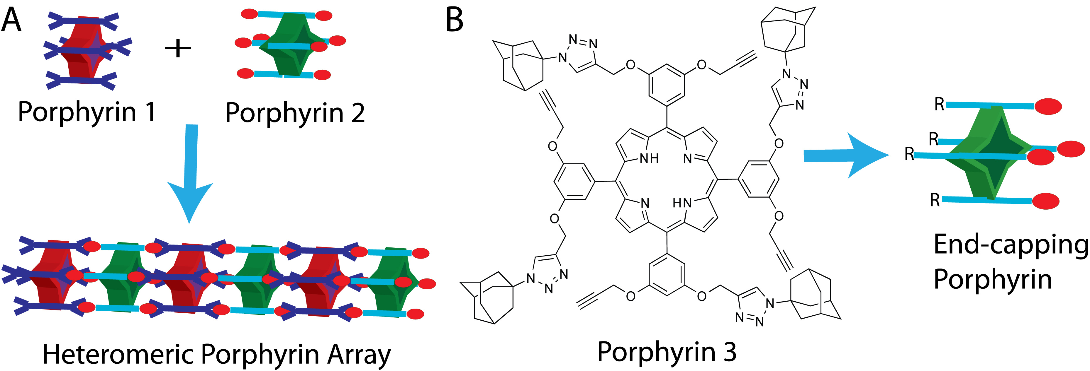
1. Results Obtained on this Project Just Prior to Commencement of ACS-PRF Funding. In preliminary results, we had demonstrated that multi-porphyrin arrays can be prepared via b-CD-fullerene interactions (Chem Comm 2009). More recently, we have also demonstrated that octadentate b-CD/adamantane interactions (i.e., the assembly of porphyrins 1 and 2, see Figure 1A) can lead to well-defined and robust multi-porphyrin arrays. These latter results were published in July 2010 (just two months prior to the commencement of the ACS-PRF funding period) and thus are not reported in the ACS PRF article submission page. Nevertheless, it is important to note that these results were of high profile since they were published in JACS as a communication (Fathalla, M.; Li, S.-C.; Neuberger, A. Schmehl, R.; Diebold, U.; Jayawickramarajah, J. Straightforward self-assembly of porphyrin nanowires in water: Harnessing adamantane/β-cyclodextrin interactions. JACS, 2010, 132, 9966-9967). Furthermore, this article was the 8th most-read article in JACS for August 2010 and was also highlighted in Nature Materials (Aug 2010, Vol. 9, p 605).
2. Progress on Trimer Self-Assembly. Based on the formation of linear porphyrin wires (that are hundreds of nanometers in length) via tandem b-CD/adamantane interactions, one of our goals was to self-assemble defined smaller trimers so that we could investigate the thermodynamic parameters of binding within a model system. Towards achieving this goal, we have just finished synthesizing tetradentate porphyrin 3 (Figure 1B) functionalized with (a) four adamantane arms and (b) also containing four alkyne arms. We are currently in the process of attaching water solubilizing PEG groups and dendrimers to porphyrin 3 via standard click chemistry procedures. After completion of the synthesis of the end-capping porphyrins, we will form self-assembled trimers (by mixing porphyrin 1 with end-capping porphyrins) and will investigate the thermodynamics of the binding process.
3a. Progress on Modulating Array Length. The tetradentate end-capping porphyrins mentioned above will also be used to modulate porphyrin array length. In particular, it is hypothesized that the oligomerization of a mixture of octa-functionalized porphyrins (e.g., 1 and 2) can be constrained by using increasing concentrations of end-capping porphyrin monomers. This controlled process should afford for us to generate shorter assemblies.
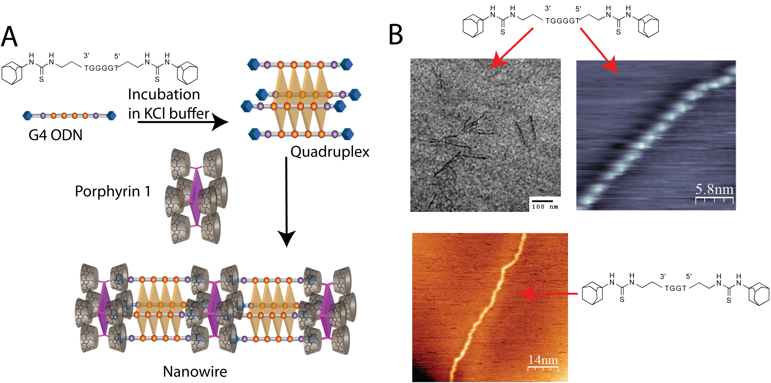
3b. Progress on Modulating Inter-Porphyrin Distances. We have designed a novel self-assembly based strategy to modulate the distance between adjacent porphyrin units within the porphyrin arrays. This straightforward strategy harnesses β-cyclodextrin/adamantane host-guest interactions in conjunction with guanine quadruplex formation. Specifically, a guanine rich oligonucleotide (ODN) strand containing adamantane head-groups assembles into a tetramolecular quadruplex in the presence of potassium cations (KCl buffer), resulting in the projection of four adamantane guests from each face (See Figure 2A). The quadruplex is then mixed with porphyrin 1 that projects four cyclodextrin hosts from each face. The resultant self-assembly has been probed via circular dichroism, STM, and TEM studies (See Figure 2B).
Importantly, the number of guanine residues on the parent ODN sequence dictates the inter-porphyrin spacing, with each guanine extending the spacing by ca. 0.34 nm. We have prepared ODNs (with adamantane head-groups) that contain 2, 4, and 6 guanines in a row. The guanine quadruplexes formed from these ODNs have been self-assembled with porphyrin 1. All the ODNs form multi-porphyrin nanowires. In addition to modulating the inter-porphyrin distance, the number of guanine residues also has an influence on the linearity of the nanowires. For instance, DNA strands with only 2 guanines form serpentine like nanoarrays with many kinks, while strands with 4 (or 6) guanines are much more straight.
4. Impact of PRF Funding on the PI's Research Program. In addition to the direct advancement of this research project, the ACS-PRF grant has supported the stipend and research activities of a graduate student, Mr. Maher Fathalla, who has now completed his PhD and is doing a post-doc at Northwestern University (with Prof. Fraser Stoddart). In addition, at the outset of this project, one of the goals of the ACS-PRF grant was to provide start-up funds that would allow our research group to obtain seminal results enabling us to become competitive for federal funding. Importantly, we have just received a National Science Foundation grant (commenced Sept/1/2011). In particular, this NSF grant, focuses on a layer-by-layer deposition method to grow vertically aligned multi-porphyrin arrays on gold and semiconductor surfaces.
Figure 1. (A) Assembly of octa-cyclodextrin functionalized porphyrin 1 in the presence of porphyrin 2. (B) Porphyrin 3 has been synthesized allowing us to potentially access tetradentate water-soluble end-capping porphyrins via click chemistry. Here R will be a water solubilizing group (e.g., PEG).
Figure 2. (A) Illustration showing the preparation of a porphyrin nanowire composed of ODNs containing 4 guanine residues. Note: The purple squares represent the porphyrin core, and light brown squares represent the G-quartets.
(B) Cryo-TEM (top left) and STM (top right) images of a porphyrin nanowire (5μM) formed from the ODN containing 4 guanines in 10 mM Tris-HCl buffer and 80 mM KCl. Note: In the Cryo-TEM image of the self-assembly of the G4 quadruplex, the observed structures have uniform dimensions with a width of ~4 nm and length between 100-150 nm. The STM image is consistent with this observation. Here, the nanowire has a width of ~2.7 nm, height of ~1.2 nm, and length > 100 nm. STM (bottom left) image of a porphyrin nanowire (5μM) formed from the ODN containing 2 guanines in 10 mM Tris-HCl buffer and 80 mM KCl. Note: The STM image of the assembly by the G2 quadruplex shows single nanowires with similar dimensions as the G4 quadruplex. However, the G2 quadruplex displays numerous kinks.


