Reports: AC9
45250-AC9 Magnetic Resonance Imaging of Oil/Water Flow through Fractures
Fractured oil reservoirs are common but immiscible fluid flow in fractures is not well understood. The standard approach has been to treat fractures as capillaries and use a Buckley-Leverett entry pressure approach to predict fluid flow. In this project, we using advanced imaging techniques to test this conceptual model of flow. We measured pressure gradients across a rock fracture as hydrocarbon and water were injected into core samples. The fluid advance was monitored using a high resolution (4.7 Tesla) magnetic resonance (MR) imaging machine designed for laboratory mice. Pressure responses were simultaneously recorded using a differential pressure transducer.
We collected samples of dolomite and sandstone for this study. However, we discovered that tracer ferric minerals in the sandstone obscured MR imaging and all fluid studies were ultimately conducted on the dolomite samples. XRD analysis indicated that the dolomite samples were almost pure CaMg(CO3)2 with small quantities of quartz and calcite. Dolomite samples were collected from large quarry pieces by drilling along natural fractures. An artificially parted sample was used as a laboratory control. This 100 mm long by 20 mm diameter solid core was cut in half, grinded and polished, and arranged such that the aperture varied linearly from 0.5 to 2 mm. The 66 mm long by 20 mm diameter naturally fractured sample presented here was parted at 2 mm by placing glass beads at the corners of the fractures. Both cores were sealed and equipped with end fittings to connect to tubing.
The fracture aperture (thickness) and mean elevation were measured using a high resolution scan of the water saturated core (Figure 1). Both the aperture and elevation vary throughout the sample. Flow experiments were conducted by forcing water through the core using a peristaltic pump. MR images were captured continuously resulting in a time-lapse analysis of immiscible fluid displacement (Figure 2). Pressure gradient was measured using a plastic pressure transducer placed inside the MR coil. Transducer mV response was monitored using wires connected to dataloggers placed outside of the MR room. The development of an effective technique for measuring hydraulic resistance in an intense magnetic field and confined space was extremely challenging and occupied a large fraction of the total project effort.
The laboratory control tests demonstrated that the pressure differential could be measured with a precision of 1 Pa. The control was also used to establish a water/dodecane/rock contact angle of 45 degrees, as measured through the water. Flow in the natural rock fracture was complex and responded to both the topography of the fracture and small aperture reductions. Capillary and buoyancy produced the same order of magnitude of pressure resistance (Figure 2). These experiments suggest that non-wetting oil may be trapped not only in larger apertures but also in small rises in topography within the fractures. Thus, Buckley-Leverett models might be modified to include both aperture distribution and topography as a pore-size distribution function.
The primary findings from this work are:
1. Dynamic measurements of immiscible flow and pressure resistance through rock fractures can be accomplished using MR imaging
2. Immiscible flow in fractures is influence by both aperture reduction and fracture topography.
3. Topographic trapping in fractures may be a significant mechanism for oil retention in fractured reservoirs.
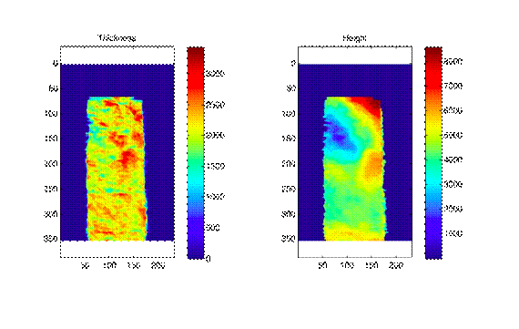
Figure 1. Fracture aperture (thickness) and mean elevation (above arbitrary datum) of the fracture measured in microns.
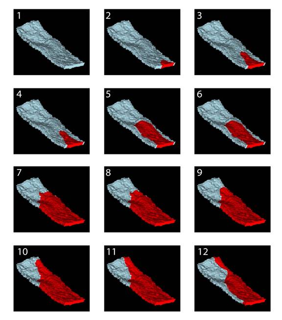
Figure 2. Dodecane advance (red) through a perspective view of the fracture void (gray).


