Reports: GB10
46693-GB10 Charge Transfer and Charge Transport in Nanofibers of Conjugated Polymer and ZnO Nanoparticles
Hybrid organic semiconductor/inorganic nanomaterials are of great interest not only as potential materials for photovoltaic applications but also as model systems for studying the fundamental physical properties and interactions at the nanometer scale. In recent years, progresses have been made in understanding the mechanism of photoinduced charge transfer process and the effect of the morphology on the charge transfer properties. However, the effects of polymer chain conformation and order/disorder in the nanocomposites on the exciton formation and dissociation, charge transfer, and charge transport properties have not been well addressed.
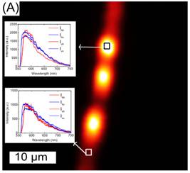
Our last report included the preparation of the conjugated polymer poly[2-methoxy-5-(2'-ethyl- hexyloxy)-1,4-phenylene vinylene] (MEH-PPV) in poly(ethylene oxide) (PEO) matrix with spincoated thin film or electrospun nanofiber morphologies and the studies of the fluorescence emission spectra of these composites with various MEH-PPV/PEO compositions. To understand the effect of local morphology on the fluorescence properties of MEH-PPV, we have measured the polarized emission spectra of a single electrospun nanofiber with MEH-PPV/PEO mass ratio of 1:10 using the user facilities of the Argonne National Laboratory. A confocal fluorescence microscope image of an individual electrospun nanofiber is shown in Figure (a). The image shows the inhomogeneous local morphology of the electrospun fibers. It is evident that the spectra from both the bright and the dimmed spots have essentially the same features, suggesting that the aggregation and chain conformation of MEH-PPV/PEO nanofibers in both bright and dimmed regions are very similar, and the observed inhomogeneity in the emission intensities is not a result of the phase separation between MEH-PPV and PEO. In addition, the polarization studies on a single nanofiber with confocal fluorescence microscope show no polarization dependence. The results have been published in Synthetic Metals.
|
|
Figure A: Confocal image of a typical MEH-PPV/PEO nanofiber; Inset shows the emission spectra of MEH-PPV with different polarization configurations at a bright spot and at a dimmed spot of the nanofiber. |
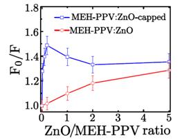
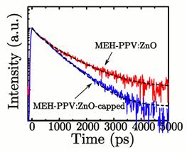
We have also been working on the surface modification of ZnO nanoparticles in order to prepare well-dispersed ZnO nanoparticle/MEH-PPV blends. ZnO nanoparticles with PVP as capping agent (ZnO-capped) are synthesized from methanol and zinc acetate dihydrate in ambient condition. The PVP capping of ZnO nanoparticles leads to better dispersion of the nanoparticles in THF solution. The fluorescence of MEH-PPV is quenched via a charge transfer process by adding ZnO or ZnO-capped nanoparticles. Compared to MEH-PPV/ZnO, steady-state Stern-Volmer plot and time-resolved measurement show that MEH-PPV is quenched more effectively and has shorter lifetimes by adding ZnO-capped nanoparticles. (Figure b) The observation is attributed to the better dispersion of ZnO-capped in MEH-PPV and the resultant larger interfacial area in MEH-PPV/ZnO-capped composites. These results conclude that the adsorption of PVP polymer on the nanoparticle surface can lead to more efficient charge transfer from conjugated polymer to nanoparticle, which may result in more efficient hybrid photovoltaic cells. Studies along this direction are underway. A manuscript based on these results has been submitted to Applied Surface Science.
|
|
|
|
Figure B: Left: Stern-Volmer plot for fluorescence quenching of MEH-PPV by ZnO or ZnO-capped in THF solution; Right: Normalized time-resolved fluorescence decays of MEH-PPV/ZnO and MEH-PPV/ZnO-capped composite films, monitored at lex=515 nm and lem>550 nm. The mass ratio of MEH-PPV to ZnO (or ZnO-capped) is 5:1. |
|
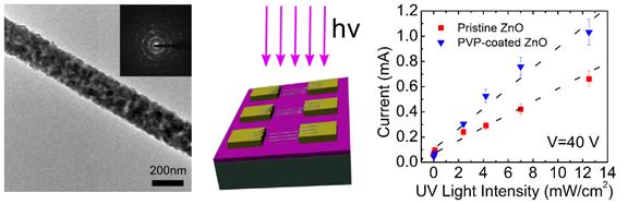
The other aspect of the project is preparation of electrospun ZnO nanofibers. We have successfully demonstrated the preparation of aligned electrospun ZnO nanofibers for simple and sensitive ultraviolet nanosensors. As shown in Figure c, the current-voltage characteristics of the nanosensors based on the electrospun ZnO nanofibers are highly sensitive to UV irradiation. The sensitivity can be further enhanced by surface coating the nanofibers with PVP. The photosensitivity is attributed to the release of the trapped electrons at the grain boundary and the surface of the nanofibers under UV irradiation. Further work on design and fabrication of highly effective chemical sensors based on the electrospun ZnO nanofibers is in progress. Our study also suggests that the electrospinning technique can be utilized as a general and simple approach to produce one-dimensional semiconducting nanomaterials which can be readily integrated into optoelectronic devices. The results have been published in Chemical Communications.
|
|
|
Figure C: Left: TEM image and electron diffraction pattern (inset) of a representative electrospun ZnO nanofiber; Middle: Schematic representation of the UV nanosensors made of the uni-axially aligned electrospun ZnO nanofibers; Right: The current measured at 40 V as a function of the UV light intensity for the two devices made of pristine ZnO and PVP-coated ZnO nanofibers, respectively. |