Reports: AC9
48505-AC9 Biosurfactant Enhanced Oil Recovery: A Pore-Scale Investigation of Interfacial and Microbial Interactions
Microbially enhanced oil recovery (MEOR) has historically been a technology marketed to industry with promises of increased tertiary oil recovery at a fraction of the price of other enhanced oil recovery processes (EOR), however to this point, increased oil production has been at best inconsistent and results of laboratory and field scale experiments and implementations have been conflicting.
The proposed work uses fundamental research approaches to evaluate MEOR technology, specifically using in-situ microbially produced biosurfactants. The research combines a novel 3D pore-scale imaging technique (high-resolution computed microtomography, CMT), 2D micro-model work, and 1D macro-scale column studies to improve understanding of the microbial and interfacial interactions at a very basic level. The focus of the proposed study is (1) effects of pore size, and pore-size distribution (to evaluate the effect of residual oil morphology and pore space geometry and thus tortuosity on mobilization), (2) effects of biosurfactant production on the wettability of the system and thus oil recovery rates, and (3) effects of inherent reservoir wettability on the effectiveness of biosurfactant-facilitated MEOR.
Research during the first year has focused on (1) the determination of optimal growth conditions for Bacillus mojavensis JF-2 and therefore maximal reductions in surface tension, (2) selection of a proper contrast agent for X-ray computed microtomography (CMT), and (3) identification of wettablity alteration in CMT images. The period of research activity by the graduate student researcher was 9 months during this first year.
(1) Optimizing Microbial Growth for Maximum Surface Tension Reduction
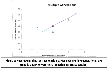
A growth medium, adapted from a mineral salts medium used by Lin et al. (1990), was altered to optimize surface tension reduction under both anaerobic and aerobic conditions by adjusting sodium chloride and potassium phosphate concentrations. However, during media optimization, progressively smaller reductions in surface tension were found over multiple generations, see Figure 1. Javaheri et al. (1985) reported this same phenomenon and suggested that lower yield biosurfactant producing isolates are selected for within the laboratory environment. It is possible that the microbe stock we received were already unintentionally pre-selected for the low yield isolate. To alleviate this difficulty, a large stock of first generation microbes were frozen and used for subsequent experiments. Once optimal media parameters were selected, baseline batch growth studies, from first generation frozen stock, were conducted for the quality control of future experiments.
Figure 1: Recorded minimal surface tension values over multiple generations, the trend is clearly towards less reduction in surface tension.
Yet, microbial growth and surface tension reduction continued to suffer under strict anaerobic conditions. To increase growth efficiency a series of tests were conducted with nitrate available as a terminal electron acceptor. When utilized by a microbe, availability of nitrate increases the amount of energy potentially extracted from a glucose molecule under anaerobic conditions. Bacillus mojavensis was unable to utilize nitrate, i.e., nitrite was not present in depleted media, and no positive growth response was apparent. Addition of yeast extract to anaerobic cultures increased microbial growth and surface tension reduction. However, complete unavailability of yeast extract to anaerobic cultures was not detrimental to growth, which conflicts with results reported by Javaheri et al. (1985). This disagreement adds to the discrepancy that already exists in the literature: Folmsbee et al. (2004) reported that anaerobic cultures must be supplemented with exogenous DNA (yeast extract is insufficient) while Javaheri et al. (1985) claims that yeast extract will support growth. Thus, one of our goals for the second year is to lift the ambiguity of the exact anaerobic growth conditions required for optimal biosurfactant production.
(2) Testing of X-Ray Contrast Agents
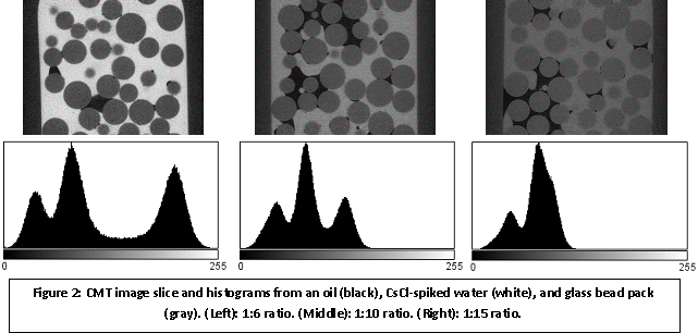
To facilitate CMT investigations glass flow-through columns (8 mm i.d.) were designed which can be scanned continuously during a water flood experiment. For the extraction of quantitative information from the gray-scale CMT data, sufficient contrast must exist between the respective phases (i.e. brine, oil, and glass). Contrast is often achieved by imaging at a monochromatic energy below and above the photoelectric edge of a given contrast agent. Three commonly used contrast agents were tested for microbial inhibition and adequate X-ray attenuation characteristics, CsCl, KI, and Iodoheptane. Cesium chloride was found to be the most benign, and was used at the Advanced Photon Source (APS), Argonne National Laboratory, to image oil water systems and determine the concentration needed for contrast. Three oil water systems at different cesium chloride concentrations were imaged at the APS, directly above the photoelectric edge of cesium at 36 keV (Figure 2). The concentrations shown are 1:6, 1:10, and 1:15 (CsCl to H2O); the data shows that only the 1:6 and 1:10 concentrations provided sufficient contrast for separation of the three phases.
(3) Identifying wettability alteration in CMT images
In direct support of our research objectives, a third task for the first year was to verify that the resolution and quality of image data were such that changes in wettability of the porous medium can be determined. Figure 2 also illustrates that it is indeed possible to detect changes in wettability of the solid phase (glass beads in this case), in this case as a result of exposure to oil (Soltrol 220) for only a couple of weeks. In the grey-scale images, one can easily detect curvatures indicative of a medium that is mixed oil- and water-wet, despite that the glass beads were water-wet when the samples were packed and saturated with the oil-water mixture. The images show that the resolution is sufficient to subsequently process such images for determination of contact angles and interfacial characteristics.
Figure 2: CMT image slice and histograms from an oil (black), CsCl-spiked water (white), and glass bead pack (gray). (Left): 1:6 ratio. (Middle): 1:10 ratio. (Right): 1:15 ratio.


